2JLP | pdb_00002jlp

Crystal structure of human extracellular copper-zinc superoxide dismutase.


Experimental Data Snapshot

  • Method: X-RAY DIFFRACTION
  • Resolution: 1.70 Å
  • R-Value Free: 
    0.185 (Depositor), 0.193 (DCC) 
  • R-Value Work: 
    0.150 (Depositor), 0.163 (DCC) 
  • R-Value Observed: 
    0.152 (Depositor) 

Starting Model: experimental
View more details

wwPDB Validation 3D Report Full Report

Validation slider image for 2JLP

This is version 1.5 of the entry. See complete history

Literature

The Structure of Human Extracellular Copper-Zinc Superoxide Dismutase at 1.7 A Resolution: Insights Into Heparin and Collagen Binding.

Antonyuk, S.V.Strange, R.W.Marklund, S.L.Hasnain, S.S.

(2009) J Mol Biology 388: 310

  • DOI: https://doi.org/10.1016/j.jmb.2009.03.026
  • Primary Citation Related Structures: 
    2JLP

  • PubMed Abstract: 

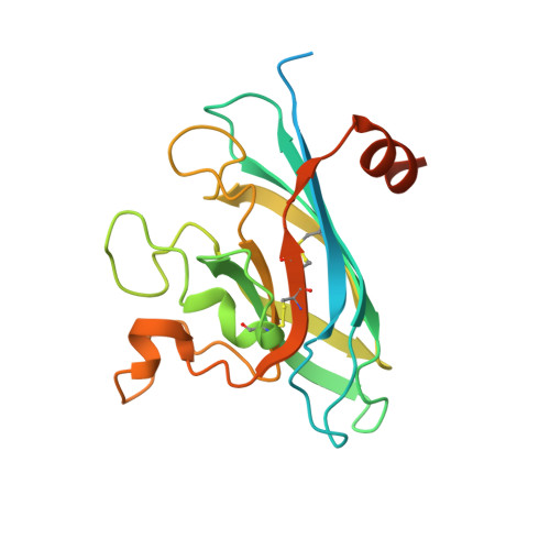
    Extracellular superoxide dismutase (SOD3) is a homotetrameric copper- and zinc-containing glycoprotein with affinity for heparin. The level of SOD3 is particularly high in blood vessel walls and in the lungs. The enzyme has multiple roles including protection of the lungs against hyperoxia and preservation of nitric oxide. The common mutation R213G, which reduces the heparin affinity of SOD3, is associated with increased risk of myocardial infarctions and stroke. We report the first crystal structure of human SOD3 at 1.7 A resolution. The overall subunit fold and the subunit-subunit interface of the SOD3 dimer are similar to the corresponding structures in Cu-Zn SOD (SOD1). The metal-binding sites are similar to those found in SOD1, but with Asn180 replacing Thr137 at the Cu-binding site and a much shorter loop at the zinc-binding site. The dimers form a functional homotetramer that is fashioned through contacts between two extended loops on each subunit. The N- and C-terminal end regions required for tetramerisation and heparin binding, respectively, are highly flexible. Two grooves fashioned by the tetramer interface are suggestive as the probable sites for heparin and collagen binding.


  • Organizational Affiliation
    • Molecular Biophysics Group, School of Biological Sciences, University of Liverpool, Liverpool L69 7ZB, UK.

Macromolecule Content 

  • Total Structure Weight: 97.42 kDa 
  • Atom Count: 5,992 
  • Modeled Residue Count: 670 
  • Deposited Residue Count: 888 
  • Unique protein chains: 1

Macromolecules

Find similar proteins by:|  3D Structure
Entity ID: 1
MoleculeChains  Sequence LengthOrganismDetailsImage
EXTRACELLULAR SUPEROXIDE DISMUTASE (CU-ZN)
A, B, C, D
222Homo sapiensMutation(s): 0 
EC: 1.15.1.1
UniProt & NIH Common Fund Data Resources
Find proteins for P08294 (Homo sapiens)
Explore P08294 
Go to UniProtKB:  P08294
PHAROS:  P08294
GTEx:  ENSG00000109610 
Entity Groups
Sequence Clusters30% Identity50% Identity70% Identity90% Identity95% Identity100% Identity
UniProt GroupP08294
Sequence Annotations
Expand
Reference Sequence

Small Molecules

Ligands 3 Unique
IDChains Name / Formula / InChI Key2D Diagram3D Interactions
ZN

Query on ZN


Download:Ideal Coordinates CCD File
F [auth A],
H [auth B],
K [auth C],
N [auth D]
ZINC ION
Zn
PTFCDOFLOPIGGS-UHFFFAOYSA-N
CU

Query on CU


Download:Ideal Coordinates CCD File
E [auth A],
G [auth B],
J [auth C],
M [auth D]
COPPER (II) ION
Cu
JPVYNHNXODAKFH-UHFFFAOYSA-N
SCN

Query on SCN


Download:Ideal Coordinates CCD File
I [auth B],
L [auth C]
THIOCYANATE ION
C N S
ZMZDMBWJUHKJPS-UHFFFAOYSA-M

Experimental Data & Validation

Experimental Data

  • Method: X-RAY DIFFRACTION
  • Resolution: 1.70 Å
  • R-Value Free:  0.185 (Depositor), 0.193 (DCC) 
  • R-Value Work:  0.150 (Depositor), 0.163 (DCC) 
  • R-Value Observed: 0.152 (Depositor) 
Space Group: P 1 21 1
Unit Cell:
Length ( Å )Angle ( ˚ )
a = 56.738α = 90
b = 93.588β = 106.23
c = 75.6γ = 90
Software Package:
Software NamePurpose
REFMACrefinement
HKL-2000data reduction
HKL-2000data scaling
MOLREPphasing

Structure Validation

View Full Validation Report


Entry History 

Deposition Data

Revision History  (Full details and data files)

  • Version 1.0: 2009-03-17
    Type: Initial release
  • Version 1.1: 2011-07-13
    Changes: Advisory, Version format compliance
  • Version 1.2: 2019-04-03
    Changes: Data collection, Other, Source and taxonomy
  • Version 1.3: 2020-03-04
    Changes: Derived calculations, Other
  • Version 1.4: 2023-12-13
    Changes: Data collection, Database references, Derived calculations, Refinement description
  • Version 1.5: 2024-11-20
    Changes: Structure summary