Effects of Phe-to-Trp mutation and fluorotryptophan incorporation on the solution structure of cardiac troponin C, and analysis of its suitability as a potential probe for in situ NMR studies
Wang, X., Mercier, P., Letourneau, P., Sykes, B.(2005) Protein Sci 14: 2447-2460
- PubMed: 16131667 Search on PubMedSearch on PubMed Central
- DOI: https://doi.org/10.1110/ps.051595805
- Primary Citation Related Structures:
2JT0, 2JT3, 2JT8, 2JTZ - PubMed Abstract:
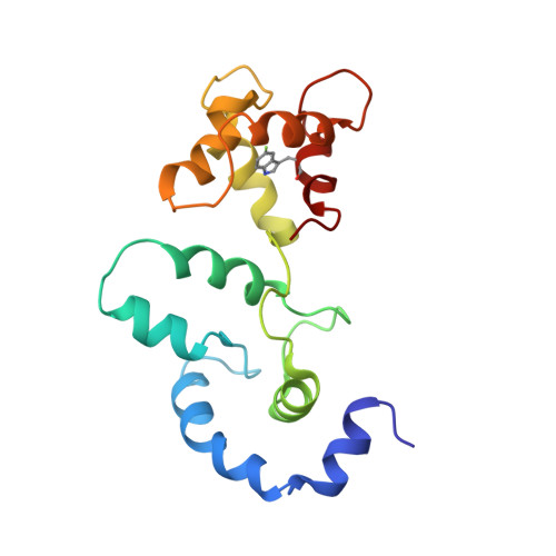
19F NMR spectroscopy is potentially a powerful tool for probing protein properties in situ. However, results obtained using this technique are relevant only if the 19F probe offers minimal perturbation to the surrounding environment. In this paper, we examine the effect of 5-fluorotryptophan (5fW) incorporation on the three-dimensional structure of cardiac troponin-C (cTnC), with the intention of developing a 19F-labeled TnC for use in in situ 19FNMR. We find that, in general, 5fW does not perturb the structure of the protein significantly. Replacement of residue Phe 153 with 5fW produces no noticeable change in protein conformation. However, replacement of residue Phe 104 with 5fW produces a folding behavior that is dependent on the Escherichia coli strain used to express the mutant. The orientations of the indole rings in these mutants are such that the Trp residue adopts a chi2 of approximately 90 degrees in the F104W mutant and approximately -100 degrees in the F153W mutant. Using results from 19F-1H heteronuclear NOE experiment, we show the replacement of L-Trp with 5fW at these positions does not change the orientation of the indole ring and the spread of the 5fW side-chain dihedral angles increases moderately for the F104(5fW) mutant and not at all for the F153(5fW) mutant. Based on these structures, we conclude that the substitution of Phe by 5fW at these two positions has minimal effects on the structure of cTnC and that the 5fW indole rings in both mutants have well defined orientation, making the two mutants viable candidates for use in in situ 19F NMR spectroscopy.
- CIHR Group in Protein Structure and Function, Department of Biochemistry, University of Alberta, Edmonton, AB, Canada T6G 2H7.
Organizational Affiliation: