2L53 | pdb_00002l53

Solution NMR Structure of apo-calmodulin in complex with the IQ motif of Human Cardiac Sodium Channel NaV1.5


Experimental Data Snapshot

  • Method: SOLUTION NMR
  • Conformers Calculated: 200 
  • Conformers Submitted: 20 
  • Selection Criteria: structures with the lowest energy 

wwPDB Validation 3D Report Full Report

Validation slider image for 2L53

This is version 1.2 of the entry. See complete history

Literature

Solution NMR Structure of Apo-Calmodulin in Complex with the IQ Motif of Human Cardiac Sodium Channel NaV1.5.

Chagot, B.Chazin, W.J.

(2011) J Mol Biology 406: 106-119

  • DOI: https://doi.org/10.1016/j.jmb.2010.11.046
  • Primary Citation Related Structures: 
    2L53

  • PubMed Abstract: 

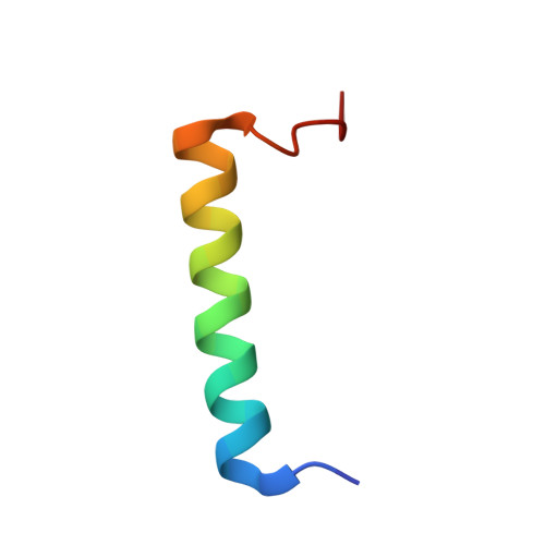
    The function of the human voltage-gated sodium channel Na(V)1.5 is regulated in part by intracellular calcium signals. The ubiquitous calcium sensor protein calmodulin (CaM) is an important part of the complex calcium-sensing apparatus in Na(V)1.5. CaM interacts with an IQ (isoleucine-glutamine) motif in the large intracellular C-terminal domain of the channel. Using co-expression and co-purification, we have been able to isolate a CaM-IQ motif complex and to determine its high-resolution structure in absence of calcium using multi-dimensional solution NMR. Under these conditions, the Na(V)1.5 IQ motif interacts with the C-terminal domain (C-lobe) of CaM, with the N-terminal domain remaining free in solution. The structure reveals that the C-lobe adopts a semi-open conformation with the IQ motif bound in a narrow hydrophobic groove. Sequence similarities between voltage-gated sodium channels and voltage-gated calcium channels suggest that the structure of the CaM-Na(V)1.5 IQ motif complex can serve as a general model for the interaction between CaM and ion channel IQ motifs under low-calcium conditions. The structure also provides insight into the biochemical basis for disease-associated mutations that map to the IQ motif in Na(V)1.5.


  • Organizational Affiliation
    • Department of Biochemistry, Vanderbilt University, Nashville, TN 37232, USA.

Macromolecule Content 

  • Total Structure Weight: 20.25 kDa 
  • Atom Count: 1,414 
  • Modeled Residue Count: 179 
  • Deposited Residue Count: 179 
  • Unique protein chains: 2

Macromolecules

Find similar proteins by:|  3D Structure
Entity ID: 1
MoleculeChains  Sequence LengthOrganismDetailsImage
Calmodulin148Homo sapiensMutation(s): 0 
Gene Names: 
CALM1CALMCAMCAM1CALM2CAM2CAMBCALM3CALML2CAM3...
CALM1CALMCAMCAM1CALM2CAM2CAMBCALM3CALML2CAM3CAMCCAMIII

Find similar proteins by:|  3D Structure
Entity ID: 2
MoleculeChains  Sequence LengthOrganismDetailsImage
Voltage-gated sodium channel type V alpha isoform b variant31Homo sapiensMutation(s): 0 
UniProt & NIH Common Fund Data Resources
Find proteins for Q14524 (Homo sapiens)
Explore Q14524 
Go to UniProtKB:  Q14524
PHAROS:  Q14524
GTEx:  ENSG00000183873 
Entity Groups
Sequence Clusters30% Identity50% Identity70% Identity90% Identity95% Identity100% Identity
UniProt GroupQ14524
Sequence Annotations
Expand
Reference Sequence

Experimental Data & Validation

Experimental Data

  • Method: SOLUTION NMR
  • Conformers Calculated: 200 
  • Conformers Submitted: 20 
  • Selection Criteria: structures with the lowest energy 

Structure Validation

View Full Validation Report


Entry History 

Deposition Data

Revision History  (Full details and data files)

  • Version 1.0: 2011-01-05
    Type: Initial release
  • Version 1.1: 2011-07-13
    Changes: Version format compliance
  • Version 1.2: 2024-05-01
    Changes: Data collection, Database references