2MV6 | pdb_00002mv6

Solution structure of the transmembrane domain and the juxta-membrane domain of the Erythropoietin Receptor in micelles


Experimental Data Snapshot

  • Method: SOLUTION NMR
  • Conformers Calculated: 50 
  • Conformers Submitted: 20 
  • Selection Criteria: structures with the lowest energy 

wwPDB Validation 3D Report Full Report

Validation slider image for 2MV6

This is version 1.1 of the entry. See complete history

Literature

Structural insight into the transmembrane domain and the juxtamembrane region of the erythropoietin receptor in micelles.

Li, Q.Wong, Y.L.Huang, Q.Kang, C.

(2014) Biophys J 107: 2325-2336

  • DOI: https://doi.org/10.1016/j.bpj.2014.10.013
  • Primary Citation Related Structures: 
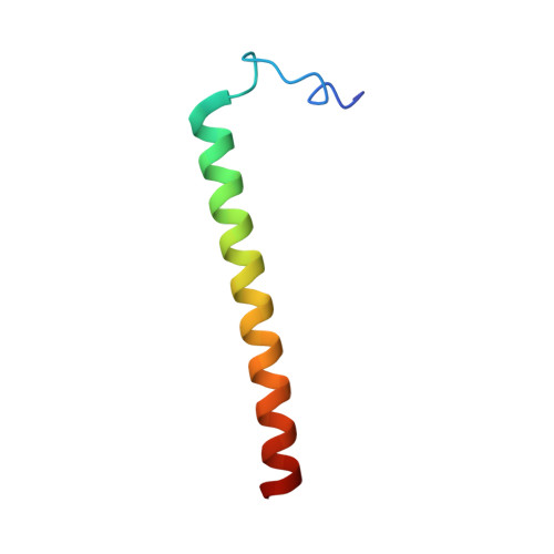
    2MV6

  • PubMed Abstract: 

    Erythropoietin receptor (EpoR) dimerization is an important step in erythrocyte formation. Its transmembrane domain (TMD) and juxtamembrane (JM) region are essential for signal transduction across the membrane. A construct compassing residues S212-P259 and containing the TMD and JM region of the human EpoR was purified and reconstituted in detergent micelles. The solution structure of the construct was determined in dodecylphosphocholine (DPC) micelles by solution NMR spectroscopy. Structural and dynamic studies demonstrated that the TMD and JM region are an ?-helix in DPC micelles, whereas residues S212-D224 at the N-terminus of the construct are not structured. The JM region is a helix that contains a hydrophobic patch formed by conserved hydrophobic residues (L253, I257, and W258). Nuclear Overhauser effect analysis, fluorescence spectroscopy, and paramagnetic relaxation enhancement experiments suggested that the JM region is exposed to the solvent. The structures of the TMD and JM region of the mouse EpoR were similar to those of the human EpoR.


  • Organizational Affiliation
    • Institute of Chemical & Engineering Sciences, Technology and Research (A(?)STAR), Singapore.

Macromolecule Content 

  • Total Structure Weight: 5.45 kDa 
  • Atom Count: 383 
  • Modeled Residue Count: 49 
  • Deposited Residue Count: 49 
  • Unique protein chains: 1

Macromolecules

Find similar proteins by:|  3D Structure
Entity ID: 1
MoleculeChains  Sequence LengthOrganismDetailsImage
Erythropoietin receptor49Homo sapiensMutation(s): 0 
Gene Names: EPOR
Membrane Entity: Yes 
UniProt & NIH Common Fund Data Resources
Find proteins for P19235 (Homo sapiens)
Explore P19235 
Go to UniProtKB:  P19235
PHAROS:  P19235
GTEx:  ENSG00000187266 
Entity Groups
Sequence Clusters30% Identity50% Identity70% Identity90% Identity95% Identity100% Identity
UniProt GroupP19235
Sequence Annotations
Expand
Reference Sequence

Experimental Data & Validation

Experimental Data

  • Method: SOLUTION NMR
  • Conformers Calculated: 50 
  • Conformers Submitted: 20 
  • Selection Criteria: structures with the lowest energy 

Structure Validation

View Full Validation Report


Entry History 

Deposition Data

Revision History  (Full details and data files)

  • Version 1.0: 2014-12-10
    Type: Initial release
  • Version 1.1: 2024-05-01
    Changes: Data collection, Database references