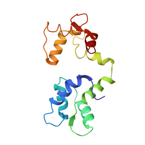
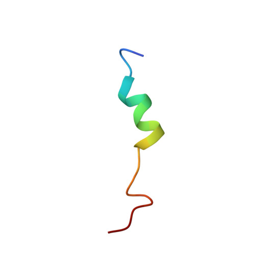
2N8J

Structure and 15N relaxation data of Calmodulin bound to the endothelial Nitric Oxide Synthase Calmodulin Binding Domain Peptide at Physiological Calcium Concentration


Experimental Data Snapshot

  • Method: SOLUTION NMR
  • Conformers Calculated: 20 
  • Conformers Submitted: 20 
  • Selection Criteria: structures with the lowest energy 

wwPDB Validation   3D Report Full Report


This is version 1.4 of the entry. See complete history


Literature

Structural Studies of a Complex Between Endothelial Nitric Oxide Synthase and Calmodulin at Physiological Calcium Concentration.

Piazza, M.Dieckmann, T.Guillemette, J.G.

(2016) Biochemistry 55: 5962-5971

  • DOI: https://doi.org/10.1021/acs.biochem.6b00821
  • Primary Citation of Related Structures:  
    2N8J

  • PubMed Abstract: 

    The small acidic protein calmodulin (CaM) serves as a Ca 2+ sensor and control element for many enzymes including nitric oxide synthase (NOS) enzymes that play major roles in key physiological and pathological processes. CaM binding causes a conformational change in NOS to allow for the electron transfer between the reductase and oxygenase domains through a process that is thought to be highly dynamic. In this report, NMR spectroscopy was used to determine the solution structure of the endothelial NOS (eNOS) peptide in complex with CaM at the lowest Ca 2+ concentration (225 nM) required for CaM to bind to eNOS and corresponds to a physiological elevated Ca 2+ level found in mammalian cells. Under these conditions, the CaM-eNOS complex has a Ca 2+ -replete C-terminal lobe bound to the eNOS peptide and a Ca 2+ free N-terminal lobe loosely associated with the eNOS peptide. With increasing Ca 2+ concentration, the binding of Ca 2+ by the N-lobe of CaM results in a stronger interaction with the C-terminal region of the eNOS peptide and increased α-helical structure of the peptide that may be part of the mechanism resulting in electron transfer from the FMN to the heme in the oxygenase domain of the enzyme. Surface plasmon resonance studies performed under the same conditions show Ca 2+ concentration-dependent binding kinetics were consistent with the NMR structural results. This investigation shows that structural studies performed under more physiological relevant conditions provide information on subtle changes in structure that may not be apparent when experiments are performed in excess Ca 2+ concentrations.


  • Organizational Affiliation

    Department of Chemistry, University of Waterloo , Waterloo, Ontario N2L 3G1, Canada.


Macromolecules
Find similar proteins by:  (by identity cutoff)  |  3D Structure
Entity ID: 1
MoleculeChains Sequence LengthOrganismDetailsImage
Calmodulin148Homo sapiensMutation(s): 0 
Gene Names: 
CALM1CALMCAMCAM1CALM2CAM2CAMBCALM3CALML2CAM3...

UniProt & NIH Common Fund Data Resources
Find proteins for P0DP23 (Homo sapiens)
Explore P0DP23 
Go to UniProtKB:  P0DP23
PHAROS:  P0DP23
GTEx:  ENSG00000198668 
Entity Groups  
Sequence Clusters30% Identity50% Identity70% Identity90% Identity95% Identity100% Identity
UniProt GroupP0DP23
Sequence Annotations
Expand
  • Reference Sequence

Find similar proteins by:  Sequence   |   3D Structure  

Entity ID: 2
MoleculeChains Sequence LengthOrganismDetailsImage
Nitric oxide synthase, endothelial22Homo sapiensMutation(s): 0 
EC: 1.14.13.39
UniProt & NIH Common Fund Data Resources
Find proteins for P29474 (Homo sapiens)
Explore P29474 
Go to UniProtKB:  P29474
PHAROS:  P29474
GTEx:  ENSG00000164867 
Entity Groups  
Sequence Clusters30% Identity50% Identity70% Identity90% Identity95% Identity100% Identity
UniProt GroupP29474
Sequence Annotations
Expand
  • Reference Sequence
Experimental Data & Validation

Experimental Data

  • Method: SOLUTION NMR
  • Conformers Calculated: 20 
  • Conformers Submitted: 20 
  • Selection Criteria: structures with the lowest energy 

Structure Validation

View Full Validation Report


Entry History 

Deposition Data

Revision History  (Full details and data files)

  • Version 1.0: 2016-10-12
    Type: Initial release
  • Version 1.1: 2017-03-15
    Changes: Database references
  • Version 1.2: 2022-08-24
    Changes: Database references
  • Version 1.3: 2023-06-14
    Changes: Other
  • Version 1.4: 2024-05-15
    Changes: Data collection, Database references