2NSU

Crystal structure of the ectodomain of human transferrin receptor fitted into a cryo-EM reconstruction of canine parvovirus and feline transferrin receptor complex


Experimental Data Snapshot

  • Method: ELECTRON MICROSCOPY
  • Resolution: 27.0 Å
  • Aggregation State: PARTICLE 
  • Reconstruction Method: SINGLE PARTICLE 

wwPDB Validation   3D Report Full Report


This is version 1.4 of the entry. See complete history


Literature

Asymmetric binding of transferrin receptor to parvovirus capsids.

Hafenstein, S.Palermo, L.M.Kostyuchenko, V.A.Xiao, C.Morais, M.C.Nelson, C.D.Bowman, V.D.Battisti, A.J.Chipman, P.R.Parrish, C.R.Rossmann, M.G.

(2007) Proc Natl Acad Sci U S A 104: 6585-6589

  • DOI: https://doi.org/10.1073/pnas.0701574104
  • Primary Citation of Related Structures:  
    2NSU

  • PubMed Abstract: 

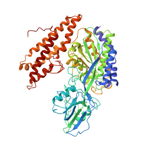
    Although many viruses are icosahedral when they initially bind to one or more receptor molecules on the cell surface, such an interaction is asymmetric, probably causing a breakdown in the symmetry and conformation of the original infecting virion in preparation for membrane penetration and release of the viral genome. Cryoelectron microscopy and biochemical analyses show that transferrin receptor, the cellular receptor for canine parvovirus, can bind to only one or a few of the 60 icosahedrally equivalent sites on the virion, indicating that either canine parvovirus has inherent asymmetry or binding of receptor induces asymmetry. The asymmetry of receptor binding to canine parvovirus is reminiscent of the special portal in tailed bacteriophages and some large, icosahedral viruses. Asymmetric interactions of icosahedral viruses with their hosts might be a more common phenomenon than previously thought and may have been obscured by averaging in previous crystallographic and electron microscopic structure determinations.


  • Organizational Affiliation

    Department of Biological Sciences, Purdue University, 915 West State Street, West Lafayette, IN 47907-2054, USA.


Macromolecules
Find similar proteins by:  (by identity cutoff)  |  3D Structure
Entity ID: 1
MoleculeChains Sequence LengthOrganismDetailsImage
Transferrin receptor protein 1
A, B
639Homo sapiensMutation(s): 1 
Gene Names: TFRC
UniProt & NIH Common Fund Data Resources
Find proteins for P02786 (Homo sapiens)
Explore P02786 
Go to UniProtKB:  P02786
PHAROS:  P02786
GTEx:  ENSG00000072274 
Entity Groups  
Sequence Clusters30% Identity50% Identity70% Identity90% Identity95% Identity100% Identity
UniProt GroupP02786
Sequence Annotations
Expand
  • Reference Sequence
Experimental Data & Validation

Experimental Data

  • Method: ELECTRON MICROSCOPY
  • Resolution: 27.0 Å
  • Aggregation State: PARTICLE 
  • Reconstruction Method: SINGLE PARTICLE 
EM Software:
TaskSoftware PackageVersion
RECONSTRUCTIONEMAN
RECONSTRUCTIONSPIDER
RECONSTRUCTIONXmipp

Structure Validation

View Full Validation Report


Entry History 

Deposition Data

Revision History  (Full details and data files)

  • Version 1.0: 2007-03-27
    Type: Initial release
  • Version 1.1: 2008-05-01
    Changes: Version format compliance
  • Version 1.2: 2011-07-13
    Changes: Version format compliance
  • Version 1.3: 2018-07-18
    Changes: Data collection
  • Version 1.4: 2023-12-27
    Changes: Data collection, Database references, Refinement description