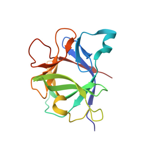
2NVH

Determination of Solvent Content in Cavities in Interleukin-1 Using Experimentally-Phased Electron Density


Experimental Data Snapshot

  • Method: X-RAY DIFFRACTION
  • Resolution: 1.53 Å
  • R-Value Observed: 0.211 

wwPDB Validation   3D Report Full Report


This is version 1.4 of the entry. See complete history


Literature

Determination of solvent content in cavities in IL-1beta using experimentally phased electron density

Quillin, M.L.Wingfield, P.T.Matthews, B.W.

(2006) Proc Natl Acad Sci U S A 103: 19749-19753

  • DOI: https://doi.org/10.1073/pnas.0609442104
  • Primary Citation of Related Structures:  
    2NVH

  • PubMed Abstract: 

    The extent to which water is present within apolar cavities in proteins remains unclear. In the case of interleukin-1beta (IL-1beta), four independent structures solved by x-ray crystallography indicate that water is not present in the central apolar cavity. In contrast, results from NMR spectroscopy suggest that water has high occupancy within the cavity but is positionally disordered, making it undetectable by standard crystallographic methods. A theoretically based crystallographic-phase refinement technique also suggested that there was the equivalent of two fully occupied water molecules within the apolar cavity. To resolve these discrepancies we sought to obtain an experimentally phased electron density map that was free of possible bias caused by mathematical modeling of the protein or the solvent. By combining native diffraction data with multiple wavelength anomalous data from a platinum derivative, accurate phases were obtained. Using these experimental phases, we estimate that occupancy of the apolar cavity in IL-1beta by solvent is close or equal to zero. Polar cavities in the protein that contain ordered solvent molecules serve as internal controls.


  • Organizational Affiliation

    Institute of Molecular Biology, Howard Hughes Medical Institute, University of Oregon, Eugene, OR 97403-1229, USA.


Macromolecules
Find similar proteins by:  (by identity cutoff)  |  3D Structure
Entity ID: 1
MoleculeChains Sequence LengthOrganismDetailsImage
Interleukin-1 beta153Homo sapiensMutation(s): 0 
Gene Names: IL1BIL1F2
UniProt & NIH Common Fund Data Resources
Find proteins for P01584 (Homo sapiens)
Explore P01584 
Go to UniProtKB:  P01584
PHAROS:  P01584
GTEx:  ENSG00000125538 
Entity Groups  
Sequence Clusters30% Identity50% Identity70% Identity90% Identity95% Identity100% Identity
UniProt GroupP01584
Sequence Annotations
Expand
  • Reference Sequence
Experimental Data & Validation

Experimental Data

  • Method: X-RAY DIFFRACTION
  • Resolution: 1.53 Å
  • R-Value Observed: 0.211 
  • Space Group: P 43
Unit Cell:
Length ( Å )Angle ( ˚ )
a = 54.506α = 90
b = 54.506β = 90
c = 75.359γ = 90
Software Package:
Software NamePurpose
DENZOdata reduction
SCALEPACKdata scaling
SHARPphasing
TNTrefinement
PDB_EXTRACTdata extraction
EPMRphasing

Structure Validation

View Full Validation Report


Entry History 

Deposition Data

Revision History  (Full details and data files)

  • Version 1.0: 2006-12-12
    Type: Initial release
  • Version 1.1: 2008-05-01
    Changes: Version format compliance
  • Version 1.2: 2011-07-13
    Changes: Version format compliance
  • Version 1.3: 2017-10-18
    Changes: Refinement description
  • Version 1.4: 2023-08-30
    Changes: Data collection, Database references, Derived calculations, Refinement description