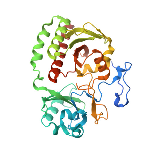
2O9C

Crystal Structure of Bacteriophytochrome chromophore binding domain at 1.45 angstrom resolution


Experimental Data Snapshot

  • Method: X-RAY DIFFRACTION
  • Resolution: 1.45 Å
  • R-Value Free: 0.192 
  • R-Value Work: 0.165 
  • R-Value Observed: 0.166 

Starting Model: experimental
View more details

wwPDB Validation   3D Report Full Report


Ligand Structure Quality Assessment 


This is version 1.9 of the entry. See complete history


Literature

High resolution structure of deinococcus bacteriophytochrome yields new insights into phytochrome architecture and evolution.

Wagner, J.R.Zhang, J.Brunzelle, J.S.Vierstra, R.D.Forest, K.T.

(2007) J Biol Chem 282: 12298-12309

  • DOI: https://doi.org/10.1074/jbc.M611824200
  • Primary Citation of Related Structures:  
    2O9B, 2O9C

  • PubMed Abstract: 

    Phytochromes are red/far red light photochromic photoreceptors that direct many photosensory behaviors in the bacterial, fungal, and plant kingdoms. They consist of an N-terminal domain that covalently binds a bilin chromophore and a C-terminal region that transmits the light signal, often through a histidine kinase relay. Using x-ray crystallography, we recently solved the first three-dimensional structure of a phytochrome, using the chromophore-binding domain of Deinococcus radiodurans bacterial phytochrome assembled with its chromophore, biliverdin IXalpha. Now, by engineering the crystallization interface, we have achieved a significantly higher resolution model. This 1.45A resolution structure helps identify an extensive buried surface between crystal symmetry mates that may promote dimerization in vivo. It also reveals that upon ligation of the C3(2) carbon of biliverdin to Cys(24), the chromophore A-ring assumes a chiral center at C2, thus becoming 2(R),3(E)-phytochromobilin, a chemistry more similar to that proposed for the attached chromophores of cyanobacterial and plant phytochromes than previously appreciated. The evolution of bacterial phytochromes to those found in cyanobacteria and higher plants must have involved greater fitness using more reduced bilins, such as phycocyanobilin, combined with a switch of the attachment site from a cysteine near the N terminus to one conserved within the cGMP phosphodiesterase/adenyl cyclase/FhlA domain. From analysis of site-directed mutants in the D. radiodurans phytochrome, we show that this bilin preference was partially driven by the change in binding site, which ultimately may have helped photosynthetic organisms optimize shade detection. Collectively, these three-dimensional structural results better clarify bilin/protein interactions and help explain how higher plant phytochromes evolved from prokaryotic progenitors.


  • Organizational Affiliation

    Departments of Genetics and Bacteriology, University of Wisconsin, Madison, Wisconsin 53706, USA.


Macromolecules
Find similar proteins by:  (by identity cutoff)  |  3D Structure
Entity ID: 1
MoleculeChains Sequence LengthOrganismDetailsImage
Bacteriophytochrome342Deinococcus radioduransMutation(s): 1 
Gene Names: bphP
EC: 2.7.13.3
UniProt
Find proteins for Q9RZA4 (Deinococcus radiodurans (strain ATCC 13939 / DSM 20539 / JCM 16871 / CCUG 27074 / LMG 4051 / NBRC 15346 / NCIMB 9279 / VKM B-1422 / R1))
Explore Q9RZA4 
Go to UniProtKB:  Q9RZA4
Entity Groups  
Sequence Clusters30% Identity50% Identity70% Identity90% Identity95% Identity100% Identity
UniProt GroupQ9RZA4
Sequence Annotations
Expand
  • Reference Sequence
Small Molecules
Ligands 1 Unique
IDChains Name / Formula / InChI Key2D Diagram3D Interactions
LBV
Query on LBV

Download Ideal Coordinates CCD File 
B [auth A]3-[2-[(Z)-[3-(2-carboxyethyl)-5-[(Z)-(4-ethenyl-3-methyl-5-oxidanylidene-pyrrol-2-ylidene)methyl]-4-methyl-pyrrol-1-ium -2-ylidene]methyl]-5-[(Z)-[(3E)-3-ethylidene-4-methyl-5-oxidanylidene-pyrrolidin-2-ylidene]methyl]-4-methyl-1H-pyrrol-3- yl]propanoic acid
C33 H37 N4 O6
DKMLMZVDTGOEGU-ISEYCTJISA-O
Experimental Data & Validation

Experimental Data

  • Method: X-RAY DIFFRACTION
  • Resolution: 1.45 Å
  • R-Value Free: 0.192 
  • R-Value Work: 0.165 
  • R-Value Observed: 0.166 
  • Space Group: C 1 2 1
Unit Cell:
Length ( Å )Angle ( ˚ )
a = 89.333α = 90
b = 51.75β = 116.29
c = 80.437γ = 90
Software Package:
Software NamePurpose
DENZOdata reduction
SCALEPACKdata scaling
AMoREphasing
REFMACrefinement
PDB_EXTRACTdata extraction
HKL-2000data collection
HKL-2000data reduction

Structure Validation

View Full Validation Report


Ligand Structure Quality Assessment 


Entry History 

Deposition Data

Revision History  (Full details and data files)

  • Version 1.0: 2007-03-06
    Type: Initial release
  • Version 1.1: 2008-05-01
    Changes: Version format compliance
  • Version 1.2: 2011-07-13
    Changes: Advisory, Derived calculations, Version format compliance
  • Version 1.3: 2011-09-28
    Changes: Atomic model
  • Version 1.4: 2017-10-18
    Changes: Refinement description
  • Version 1.5: 2018-01-24
    Changes: Structure summary
  • Version 1.6: 2021-10-20
    Changes: Database references, Derived calculations, Structure summary
  • Version 1.7: 2023-08-30
    Changes: Data collection, Refinement description
  • Version 1.8: 2024-03-13
    Changes: Source and taxonomy, Structure summary
  • Version 1.9: 2024-10-30
    Changes: Structure summary