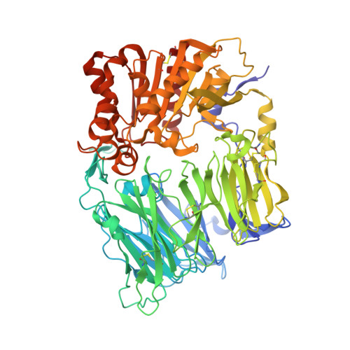
2OAG

Crystal structure of human dipeptidyl peptidase IV (DPPIV) with pyrrolidine-constrained phenethylamine 29g


Experimental Data Snapshot

  • Method: X-RAY DIFFRACTION
  • Resolution: 2.30 Å
  • R-Value Free: 0.240 
  • R-Value Work: 0.197 

wwPDB Validation   3D Report Full Report


Ligand Structure Quality Assessment 


This is version 1.3 of the entry. See complete history


Literature

Pyrrolidine-constrained phenethylamines: The design of potent, selective, and pharmacologically efficacious dipeptidyl peptidase IV (DPP4) inhibitors from a lead-like screening hit.

Backes, B.J.Longenecker, K.Hamilton, G.L.Stewart, K.Lai, C.Kopecka, H.von Geldern, T.W.Madar, D.J.Pei, Z.Lubben, T.H.Zinker, B.A.Tian, Z.Ballaron, S.J.Stashko, M.A.Mika, A.K.Beno, D.W.Kempf-Grote, A.J.Black-Schaefer, C.Sham, H.L.Trevillyan, J.M.

(2007) Bioorg Med Chem Lett 17: 2005-2012

  • DOI: https://doi.org/10.1016/j.bmcl.2007.01.026
  • Primary Citation of Related Structures:  
    2OAE, 2OAG

  • PubMed Abstract: 

    A novel series of pyrrolidine-constrained phenethylamines were developed as dipeptidyl peptidase IV (DPP4) inhibitors for the treatment of type 2 diabetes. The cyclohexene ring of lead-like screening hit 5 was replaced with a pyrrolidine to enable parallel chemistry, and protein co-crystal structural data guided the optimization of N-substituents. Employing this strategy, a >400x improvement in potency over the initial hit was realized in rapid fashion. Optimized compounds are potent and selective inhibitors with excellent pharmacokinetic profiles. Compound 30 was efficacious in vivo, lowering blood glucose in ZDF rats that were allowed to feed freely on a mixed meal.


  • Organizational Affiliation

    Metabolic Disease Research, Abbott Laboratories, Abbott Park Road, Abbott Park, IL 60064-6099, USA. bradley.backes@abbott.com


Macromolecules
Find similar proteins by:  (by identity cutoff)  |  3D Structure
Entity ID: 1
MoleculeChains Sequence LengthOrganismDetailsImage
Dipeptidyl peptidase 4
A, B, C, D
726Homo sapiensMutation(s): 0 
Gene Names: DPP4ADCP2CD26
EC: 3.4.14.5
UniProt & NIH Common Fund Data Resources
Find proteins for P27487 (Homo sapiens)
Explore P27487 
Go to UniProtKB:  P27487
PHAROS:  P27487
GTEx:  ENSG00000197635 
Entity Groups  
Sequence Clusters30% Identity50% Identity70% Identity90% Identity95% Identity100% Identity
UniProt GroupP27487
Sequence Annotations
Expand
  • Reference Sequence
Small Molecules
Ligands 1 Unique
IDChains Name / Formula / InChI Key2D Diagram3D Interactions
DLI
Query on DLI

Download Ideal Coordinates CCD File 
E [auth B](3R,4S)-1-{6-[3-(METHYLSULFONYL)PHENYL]PYRIMIDIN-4-YL}-4-(2,4,5-TRIFLUOROPHENYL)PYRROLIDIN-3-AMINE
C21 H19 F3 N4 O2 S
OAWGQHXWGXOUKV-BEFAXECRSA-N
Binding Affinity Annotations 
IDSourceBinding Affinity
DLI PDBBind:  2OAG Ki: 3.4 (nM) from 1 assay(s)
Experimental Data & Validation

Experimental Data

  • Method: X-RAY DIFFRACTION
  • Resolution: 2.30 Å
  • R-Value Free: 0.240 
  • R-Value Work: 0.197 
  • Space Group: P 1 21 1
Unit Cell:
Length ( Å )Angle ( ˚ )
a = 120.006α = 90
b = 126.225β = 96.51
c = 127.563γ = 90
Software Package:
Software NamePurpose
DENZOdata reduction
SCALEPACKdata scaling
CNSrefinement
PDB_EXTRACTdata extraction
HKL-2000data collection
MOLREPphasing

Structure Validation

View Full Validation Report


Ligand Structure Quality Assessment 


Entry History 

Deposition Data

Revision History  (Full details and data files)

  • Version 1.0: 2007-09-11
    Type: Initial release
  • Version 1.1: 2011-07-13
    Changes: Atomic model, Database references, Derived calculations, Non-polymer description, Structure summary, Version format compliance
  • Version 1.2: 2017-10-18
    Changes: Refinement description
  • Version 1.3: 2023-12-27
    Changes: Data collection, Database references, Derived calculations