2OO0

A structural insight into the inhibition of human and Leishmania donovani ornithine decarboxylases by 3-aminooxy-1-aminopropane


Experimental Data Snapshot

  • Method: X-RAY DIFFRACTION
  • Resolution: 1.90 Å
  • R-Value Free: 0.213 
  • R-Value Work: 0.184 
  • R-Value Observed: 0.187 

wwPDB Validation   3D Report Full Report


Ligand Structure Quality Assessment 


This is version 1.4 of the entry. See complete history


Literature

A structural insight into the inhibition of human and Leishmania donovani ornithine decarboxylases by 1-amino-oxy-3-aminopropane.

Dufe, V.T.Ingner, D.Heby, O.Khomutov, A.R.Persson, L.Al-Karadaghi, S.

(2007) Biochem J 405: 261-268

  • DOI: https://doi.org/10.1042/BJ20070188
  • Primary Citation of Related Structures:  
    2ON3, 2OO0

  • PubMed Abstract: 

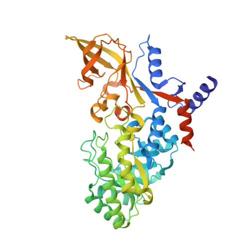
    The critical role of polyamines in key processes such as cell growth, differentiation and macromolecular synthesis makes the enzymes involved in their synthesis potential targets in the treatment of certain types of cancer and parasitic diseases. Here we present a study on the inhibition of human and Leishmania donovani ODC (ornithine decarboxylase), the first committed enzyme in the polyamine biosynthesis pathway, by APA (1-amino-oxy-3-aminopropane). The present study shows APA to be a potent inhibitor of both human and L. donovani ODC with a K(i) value of around 1.0 nM. We also show that L. donovani ODC binds the substrate, the co-enzyme pyridoxal 5'-phosphate and the irreversible inhibitor alpha-difluoromethylornithine (a curative agent of West African sleeping sickness) with less affinity than human ODC. We have also determined the three-dimensional structure of human ODC in complex with APA, which revealed the mode of the inhibitor binding to the enzyme. In contrast with earlier reports, the structure showed no indication of oxime formation between APA and PLP (pyridoxal 5'-phosphate). Homology modelling suggests a similar mode of binding of APA to L. donovani ODC. A comparison of the ODC-APA-PLP structure with earlier ODC structures also shows that the protease-sensitive loop (residues 158-168) undergoes a large conformational change and covers the active site of the protein. The understanding of the structural mode of APA binding may constitute the basis for the development of more specific inhibitors of L. donovani ODC.


  • Organizational Affiliation

    Department of Molecular Biophysics, Lund University, S-221 00 Lund, Sweden.


Macromolecules
Find similar proteins by:  (by identity cutoff)  |  3D Structure
Entity ID: 1
MoleculeChains Sequence LengthOrganismDetailsImage
Ornithine decarboxylase
A, B
471Homo sapiensMutation(s): 0 
Gene Names: odc
EC: 4.1.1.17
UniProt & NIH Common Fund Data Resources
Find proteins for P11926 (Homo sapiens)
Explore P11926 
Go to UniProtKB:  P11926
PHAROS:  P11926
GTEx:  ENSG00000115758 
Entity Groups  
Sequence Clusters30% Identity50% Identity70% Identity90% Identity95% Identity100% Identity
UniProt GroupP11926
Sequence Annotations
Expand
  • Reference Sequence
Small Molecules
Ligands 4 Unique
IDChains Name / Formula / InChI Key2D Diagram3D Interactions
PLP
Query on PLP

Download Ideal Coordinates CCD File 
E [auth A],
I [auth B]
PYRIDOXAL-5'-PHOSPHATE
C8 H10 N O6 P
NGVDGCNFYWLIFO-UHFFFAOYSA-N
N2P
Query on N2P

Download Ideal Coordinates CCD File 
G [auth A],
K [auth B]
PENTANE-1,5-DIAMINE
C5 H14 N2
VHRGRCVQAFMJIZ-UHFFFAOYSA-N
XAP
Query on XAP

Download Ideal Coordinates CCD File 
F [auth A],
J [auth B]
3-AMINOOXY-1-AMINOPROPANE
C3 H10 N2 O
VSZFWDPIWSPZON-UHFFFAOYSA-N
ACT
Query on ACT

Download Ideal Coordinates CCD File 
C [auth A],
D [auth A],
H [auth B]
ACETATE ION
C2 H3 O2
QTBSBXVTEAMEQO-UHFFFAOYSA-M
Binding Affinity Annotations 
IDSourceBinding Affinity
XAP Binding MOAD:  2OO0 Ki: 1.4 (nM) from 1 assay(s)
BindingDB:  2OO0 IC50: 35 (nM) from 1 assay(s)
PLP Binding MOAD:  2OO0 Kd: 50 (nM) from 1 assay(s)
Experimental Data & Validation

Experimental Data

  • Method: X-RAY DIFFRACTION
  • Resolution: 1.90 Å
  • R-Value Free: 0.213 
  • R-Value Work: 0.184 
  • R-Value Observed: 0.187 
  • Space Group: C 1 2 1
Unit Cell:
Length ( Å )Angle ( ˚ )
a = 108.018α = 90
b = 87.11β = 91.02
c = 130.08γ = 90
Software Package:
Software NamePurpose
REFMACrefinement
MAR345data collection
XDSdata reduction
XSCALEdata scaling
PHASERphasing

Structure Validation

View Full Validation Report


Ligand Structure Quality Assessment 


Entry History 

Deposition Data

Revision History  (Full details and data files)

  • Version 1.0: 2007-07-17
    Type: Initial release
  • Version 1.1: 2008-05-01
    Changes: Version format compliance
  • Version 1.2: 2011-07-13
    Changes: Non-polymer description, Version format compliance
  • Version 1.3: 2017-10-18
    Changes: Data collection, Refinement description
  • Version 1.4: 2023-08-30
    Changes: Data collection, Database references, Derived calculations, Refinement description