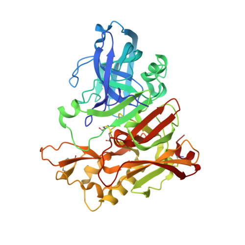
2P4J

Crystal structure of beta-secretase bond to an inhibitor with Isophthalamide Derivatives at P2-P3


Experimental Data Snapshot

  • Method: X-RAY DIFFRACTION
  • Resolution: 2.50 Å
  • R-Value Free: 0.259 
  • R-Value Work: 0.201 
  • R-Value Observed: 0.201 

Starting Model: experimental
View more details

wwPDB Validation   3D Report Full Report


Ligand Structure Quality Assessment 

Created with Raphaël 2.3.0Worse 01 BetterLigand structure goodness of fit to experimental dataBest fitted 23IClick on this verticalbar to view details

This is version 1.5 of the entry. See complete history


Literature

Design, synthesis, and X-ray structure of potent memapsin 2 (beta-secretase) inhibitors with isophthalamide derivatives as the P2-P3-ligands.

Ghosh, A.K.Kumaragurubaran, N.Hong, L.Kulkarni, S.S.Xu, X.Chang, W.Weerasena, V.Turner, R.Koelsch, G.Bilcer, G.Tang, J.

(2007) J Med Chem 50: 2399-2407

  • DOI: https://doi.org/10.1021/jm061338s
  • Primary Citation of Related Structures:  
    2P4J

  • PubMed Abstract: 

    Structure-based design and synthesis of a number of potent and selective memapsin 2 inhibitors are described. These inhibitors were designed based upon the X-ray structure of memapsin 2-bound inhibitor 3 that incorporates methylsulfonyl alanine as the P2-ligand and a substituted pyrazole as the P3-ligand. Of particular importance, we examined the ability of the substituted isophthalic acid amide derivative to mimic the key interactions in the S2-S3 regions of the enzyme active sites of 3-bound memapsin 2. We investigated various substituted phenylethyl, alpha-methylbenzyl, and oxazolylmethyl groups as the P3-ligands. A number of inhibitors exhibited very potent inhibitory activity against mempasin 2 and good selectivity against memapsin 1. Inhibitor 5d has shown low nanomolar enzyme inhibitory potency (Ki=1.1 nM) and very good cellular inhibitory activity (IC50=39 nM). Furthermore, in a preliminary study, inhibitor 5d has shown 30% reduction of Abeta40 production in transgenic mice after a single intraperitoneal administration (8 mg/kg). A protein-ligand X-ray crystal structure of 5d-bound memapsin 2 provided vital molecular insight that can serve as an important guide to further design of novel inhibitors.


  • Organizational Affiliation

    Department of Chemistry, Purdue University, West Lafayette, Indiana 47907, USA. akghosh@purdue.edu


Macromolecules
Find similar proteins by:  (by identity cutoff)  |  3D Structure
Entity ID: 1
MoleculeChains Sequence LengthOrganismDetailsImage
Beta-secretase 1
A, B, C, D
389Homo sapiensMutation(s): 0 
Gene Names: BACE1BACE
EC: 3.4.23.46
UniProt & NIH Common Fund Data Resources
Find proteins for P56817 (Homo sapiens)
Explore P56817 
Go to UniProtKB:  P56817
PHAROS:  P56817
GTEx:  ENSG00000186318 
Entity Groups  
Sequence Clusters30% Identity50% Identity70% Identity90% Identity95% Identity100% Identity
UniProt GroupP56817
Sequence Annotations
Expand
  • Reference Sequence
Small Molecules
Binding Affinity Annotations 
IDSourceBinding Affinity
23I PDBBind:  2P4J Ki: 1.1 (nM) from 1 assay(s)
Experimental Data & Validation

Experimental Data

  • Method: X-RAY DIFFRACTION
  • Resolution: 2.50 Å
  • R-Value Free: 0.259 
  • R-Value Work: 0.201 
  • R-Value Observed: 0.201 
  • Space Group: P 1 21 1
Unit Cell:
Length ( Å )Angle ( ˚ )
a = 86.929α = 90
b = 129.969β = 97.48
c = 87.483γ = 90
Software Package:
Software NamePurpose
DENZOdata reduction
SCALEPACKdata scaling
CNSrefinement
PDB_EXTRACTdata extraction
MAR345dtbdata collection
CNSphasing

Structure Validation

View Full Validation Report


Ligand Structure Quality Assessment 

Created with Raphaël 2.3.0Worse 01 BetterLigand structure goodness of fit to experimental dataBest fitted 23IClick on this verticalbar to view details

Entry History 

Deposition Data

Revision History  (Full details and data files)

  • Version 1.0: 2007-07-24
    Type: Initial release
  • Version 1.1: 2008-05-01
    Changes: Version format compliance
  • Version 1.2: 2011-07-13
    Changes: Version format compliance
  • Version 1.3: 2017-10-18
    Changes: Refinement description
  • Version 1.4: 2023-08-30
    Changes: Data collection, Database references, Refinement description
  • Version 1.5: 2024-11-13
    Changes: Structure summary