2P5W | pdb_00002p5w

Crystal structures of high affinity human T-cell receptors bound to pMHC reveal native diagonal binding geometry


Experimental Data Snapshot

  • Method: X-RAY DIFFRACTION
  • Resolution: 2.20 Å
  • R-Value Free: 
    0.245 (Depositor), 0.245 (DCC) 
  • R-Value Work: 
    0.165 (Depositor), 0.169 (DCC) 
  • R-Value Observed: 
    0.169 (Depositor) 

Starting Model: experimental
View more details

wwPDB Validation 3D Report Full Report

Validation slider image for 2P5W

This is version 1.4 of the entry. See complete history

Literature

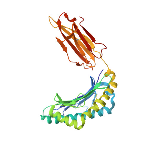
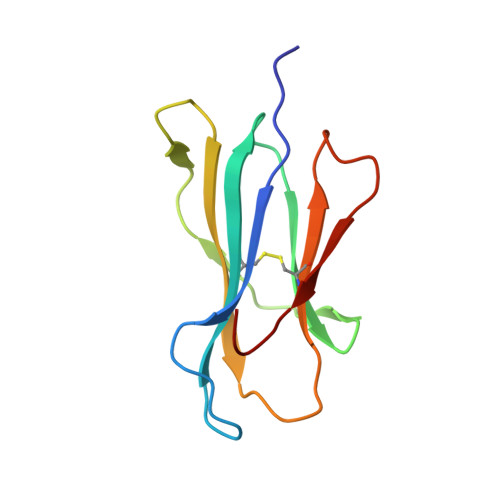
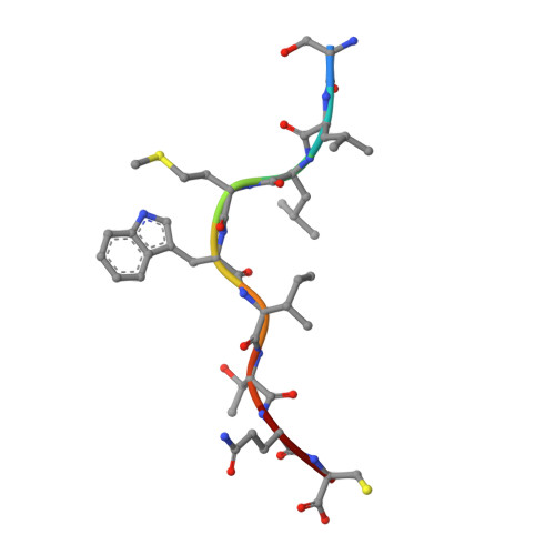
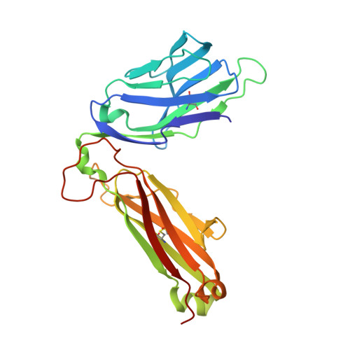
Crystal structures of high affinity human T-cell receptors bound to peptide major histocompatibility complex reveal native diagonal binding geometry

Sami, M.Rizkallah, P.J.Dunn, S.Molloy, P.Moysey, R.Vuidepot, A.Baston, E.Todorov, P.Yi, L.Gao, F.Boulter, J.M.Jakobsen, B.K.

(2007) Protein Eng Des Sel 20: 397-403

  • DOI: https://doi.org/10.1093/protein/gzm033
  • Primary Citation Related Structures: 
    2P5E, 2P5W, 2PYE, 2PYF

  • PubMed Abstract: 

    Naturally selected T-cell receptors (TCRs) are characterised by low binding affinities, typically in the range 1-100 microM. Crystal structures of syngeneic TCRs bound to peptide major histocompatibility complex (pMHC) antigens exhibit a conserved mode of binding characterised by a distinct diagonal binding geometry, with poor shape complementarity (SC) between receptor and ligand. Here, we report the structures of three in vitro affinity enhanced TCRs that recognise the pMHC tumour epitope NY-ESO(157-165) (SLLMWITQC). These crystal structures reveal that the docking mode for the high affinity TCRs is identical to that reported for the parental wild-type TCR, with only subtle changes in the mutated complementarity determining regions (CDRs) that form contacts with pMHC; both CDR2 and CDR3 mutations act synergistically to improve the overall affinity. Comparison of free and bound TCR structures for both wild-type and a CDR3 mutant reveal an induced fit mechanism arising from restructuring of CDR3 loops which allows better peptide binding. Overall, an increased interface area, improved SC and additional H-bonding interactions are observed, accounting for the increase in affinity. Most notably, there is a marked increase in the SC for the central methionine and tryptophan peptide motif over the native TCR.


  • Organizational Affiliation
    • Avidex Limited (subsidiary of Medigene Ag), 57c Milton Park, Abingdon, Oxon OX14 4RX, UK.

Macromolecule Content 

  • Total Structure Weight: 95.23 kDa 
  • Atom Count: 7,417 
  • Modeled Residue Count: 819 
  • Deposited Residue Count: 820 
  • Unique protein chains: 5

Macromolecules

Find similar proteins by:|  3D Structure
Entity ID: 1
MoleculeChains  Sequence LengthOrganismDetailsImage
HLA class I histocompatibility antigen, A-2 alpha chain276Homo sapiensMutation(s): 0 
Gene Names: HLA-AHLAA
UniProt & NIH Common Fund Data Resources
Find proteins for P04439 (Homo sapiens)
Explore P04439 
Go to UniProtKB:  P04439
PHAROS:  P04439
GTEx:  ENSG00000206503 
Entity Groups
Sequence Clusters30% Identity50% Identity70% Identity90% Identity95% Identity100% Identity
UniProt GroupP04439
Sequence Annotations
Expand
Reference Sequence
Find similar proteins by:|  3D Structure
Entity ID: 2
MoleculeChains  Sequence LengthOrganismDetailsImage
Beta-2-microglobulin100Homo sapiensMutation(s): 0 
Gene Names: B2M
UniProt & NIH Common Fund Data Resources
Find proteins for P61769 (Homo sapiens)
Explore P61769 
Go to UniProtKB:  P61769
PHAROS:  P61769
GTEx:  ENSG00000166710 
Entity Groups
Sequence Clusters30% Identity50% Identity70% Identity90% Identity95% Identity100% Identity
UniProt GroupP61769
Sequence Annotations
Expand
Reference Sequence
Find similar proteins by:  Sequence   |   3D Structure  
Entity ID: 3
MoleculeChains  Sequence LengthOrganismDetailsImage
Cancer/testis antigen 1B9N/AMutation(s): 0 
UniProt & NIH Common Fund Data Resources
Find proteins for P78358 (Homo sapiens)
Explore P78358 
Go to UniProtKB:  P78358
Entity Groups
UniProt GroupP78358
Sequence Annotations
Expand
Reference Sequence
Find similar proteins by:|  3D Structure
Entity ID: 4
MoleculeChains  Sequence LengthOrganismDetailsImage
T-CELL RECEPTOR, ALPHA CHAIN192Homo sapiensMutation(s): 0 
UniProt & NIH Common Fund Data Resources
Find proteins for P01848 (Homo sapiens)
Explore P01848 
Go to UniProtKB:  P01848
PHAROS:  P01848
Entity Groups
Sequence Clusters30% Identity50% Identity70% Identity90% Identity95% Identity100% Identity
UniProt GroupP01848
Sequence Annotations
Expand
Reference Sequence
Find similar proteins by:|  3D Structure
Entity ID: 5
MoleculeChains  Sequence LengthOrganismDetailsImage
Hypothetical protein243Homo sapiensMutation(s): 0 

Small Molecules

Ligands 5 Unique
IDChains Name / Formula / InChI Key2D Diagram3D Interactions
EPE

Query on EPE


Download:Ideal Coordinates CCD File
X [auth E]4-(2-HYDROXYETHYL)-1-PIPERAZINE ETHANESULFONIC ACID
C8 H18 N2 O4 S
JKMHFZQWWAIEOD-UHFFFAOYSA-N
SO4

Query on SO4


Download:Ideal Coordinates CCD File
F [auth A]
G [auth A]
H [auth A]
O [auth B]
R [auth D]
F [auth A],
G [auth A],
H [auth A],
O [auth B],
R [auth D],
W [auth E]
SULFATE ION
O4 S
QAOWNCQODCNURD-UHFFFAOYSA-L
GOL

Query on GOL


Download:Ideal Coordinates CCD File
I [auth A]
J [auth A]
K [auth A]
L [auth A]
P [auth B]
I [auth A],
J [auth A],
K [auth A],
L [auth A],
P [auth B],
Q [auth B],
S [auth D],
T [auth D],
U [auth D],
V [auth D],
Y [auth E]
GLYCEROL
C3 H8 O3
PEDCQBHIVMGVHV-UHFFFAOYSA-N
CA

Query on CA


Download:Ideal Coordinates CCD File
M [auth B]CALCIUM ION
Ca
BHPQYMZQTOCNFJ-UHFFFAOYSA-N
MG

Query on MG


Download:Ideal Coordinates CCD File
N [auth B]MAGNESIUM ION
Mg
JLVVSXFLKOJNIY-UHFFFAOYSA-N

Experimental Data & Validation

Experimental Data

  • Method: X-RAY DIFFRACTION
  • Resolution: 2.20 Å
  • R-Value Free:  0.245 (Depositor), 0.245 (DCC) 
  • R-Value Work:  0.165 (Depositor), 0.169 (DCC) 
  • R-Value Observed: 0.169 (Depositor) 
Space Group: P 1 21 1
Unit Cell:
Length ( Å )Angle ( ˚ )
a = 74.59α = 90
b = 52.325β = 98.13
c = 118.932γ = 90
Software Package:
Software NamePurpose
SCALAdata scaling
AMoREphasing
REFMACrefinement
PDB_EXTRACTdata extraction
MOSFLMdata reduction

Structure Validation

View Full Validation Report


Entry History 

Deposition Data

Revision History  (Full details and data files)

  • Version 1.0: 2007-09-25
    Type: Initial release
  • Version 1.1: 2011-07-13
    Changes: Advisory, Version format compliance
  • Version 1.2: 2023-08-30
    Changes: Data collection, Database references, Derived calculations, Refinement description
  • Version 1.3: 2023-12-27
    Changes: Derived calculations
  • Version 1.4: 2024-11-13
    Changes: Structure summary