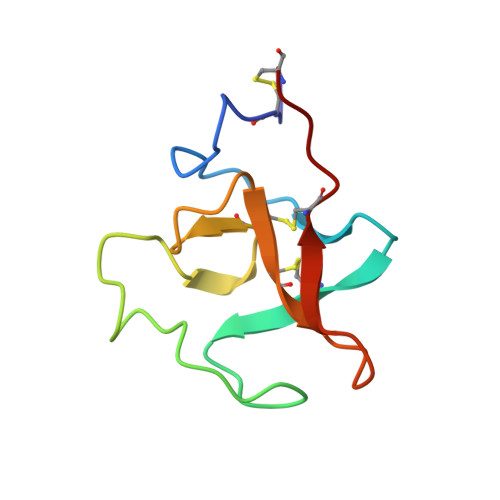
2PK4

THE REFINED STRUCTURE OF THE EPSILON-AMINOCAPROIC ACID COMPLEX OF HUMAN PLASMINOGEN KRINGLE


Experimental Data Snapshot

  • Method: X-RAY DIFFRACTION
  • Resolution: 2.25 Å
  • R-Value Observed: 0.148 

wwPDB Validation   3D Report Full Report


This is version 2.1 of the entry. See complete history


Literature

The refined structure of the epsilon-aminocaproic acid complex of human plasminogen kringle 4.

Wu, T.P.Padmanabhan, K.Tulinsky, A.Mulichak, A.M.

(1991) Biochemistry 30: 10589-10594

  • DOI: https://doi.org/10.1021/bi00107a030
  • Primary Citation of Related Structures:  
    2PK4

  • PubMed Abstract: 

    The crystallographic structure of the plasminogen kringle 4-epsilon-aminocaproic acid (ACA) complex (K4-ACA) has been solved by molecular replacement rotation-translation methods utilizing the refined apo-K4 structure as a search model (Mulichak et al., 1991), and it has been refined to an R value of 0.148 at 2.25-A resolution. The K4-ACA structure consists of two interkringle residues, the kringle along with the ACA ligand, and 106 water molecules. The lysine-binding site has been confirmed to be a relatively open and shallow depression, lined by aromatic rings of Trp62, Phe64, and Trp72, which provide a highly nonpolar environment between doubly charged anionic and cationic centers formed by Asp55/Asp57 and Lys35/Arg71. A zwitterionic ACA ligand molecule is held by hydrogen-bonded ion pair interactions and van der Waals contacts between the charged centers. The lysine-binding site of apo-K4 and K4-ACA have been compared: the rms differences in main-chain and side-chain positions are 0.25 and 0.69 A, respectively, both practically within error of the determinations. The largest deviations in the binding site are due to different crystal packing interactions. Thus, the lysine-binding site appears to be preformed, and lysine binding does not require conformational changes of the host. The results of NMR studies of lysine binding with K4 are correlated with the structure of K4-ACA and agree well.


  • Organizational Affiliation

    Department of Chemistry, Michigan State University, East Lansing 48824.


Macromolecules
Find similar proteins by:  (by identity cutoff)  |  3D Structure
Entity ID: 1
MoleculeChains Sequence LengthOrganismDetailsImage
HUMAN PLASMINOGEN KRINGLE 480Homo sapiensMutation(s): 0 
EC: 3.4.21.7
UniProt & NIH Common Fund Data Resources
Find proteins for P00747 (Homo sapiens)
Explore P00747 
Go to UniProtKB:  P00747
PHAROS:  P00747
GTEx:  ENSG00000122194 
Entity Groups  
Sequence Clusters30% Identity50% Identity70% Identity90% Identity95% Identity100% Identity
UniProt GroupP00747
Sequence Annotations
Expand
  • Reference Sequence
Small Molecules
Ligands 1 Unique
IDChains Name / Formula / InChI Key2D Diagram3D Interactions
ACA
Query on ACA

Download Ideal Coordinates CCD File 
B [auth A]6-AMINOHEXANOIC ACID
C6 H13 N O2
SLXKOJJOQWFEFD-UHFFFAOYSA-N
Binding Affinity Annotations 
IDSourceBinding Affinity
ACA BindingDB:  2PK4 Ki: 5.30e+7 (nM) from 1 assay(s)
Kd: 9000 (nM) from 1 assay(s)
IC50: min: 4.00e+4, max: 1.05e+5 (nM) from 2 assay(s)
Experimental Data & Validation

Experimental Data

  • Method: X-RAY DIFFRACTION
  • Resolution: 2.25 Å
  • R-Value Observed: 0.148 
  • Space Group: P 1 21 1
Unit Cell:
Length ( Å )Angle ( ˚ )
a = 42.21α = 90
b = 35.46β = 102.94
c = 25.43γ = 90
Software Package:
Software NamePurpose
PROFFTrefinement

Structure Validation

View Full Validation Report


Entry History 

Deposition Data

Revision History  (Full details and data files)

  • Version 1.0: 1993-10-31
    Type: Initial release
  • Version 1.1: 2008-03-03
    Changes: Version format compliance
  • Version 1.2: 2011-07-13
    Changes: Version format compliance
  • Version 1.3: 2017-11-29
    Changes: Derived calculations, Other
  • Version 2.0: 2023-11-15
    Changes: Advisory, Atomic model, Data collection, Database references, Derived calculations
  • Version 2.1: 2024-10-30
    Changes: Structure summary