2Q7N

Crystal structure of Leukemia inhibitory factor in complex with LIF receptor (domains 1-5)


Experimental Data Snapshot

  • Method: X-RAY DIFFRACTION
  • Resolution: 4.00 Å
  • R-Value Free: 0.287 
  • R-Value Work: 0.237 
  • R-Value Observed: 0.240 

wwPDB Validation   3D Report Full Report


Ligand Structure Quality Assessment 


This is version 3.1 of the entry. See complete history


Literature

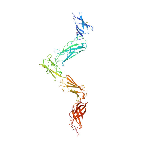
An unusual cytokine:Ig-domain interaction revealed in the crystal structure of leukemia inhibitory factor (LIF) in complex with the LIF receptor.

Huyton, T.Zhang, J.G.Luo, C.S.Lou, M.Z.Hilton, D.J.Nicola, N.A.Garrett, T.P.

(2007) Proc Natl Acad Sci U S A 104: 12737-12742

  • DOI: https://doi.org/10.1073/pnas.0705577104
  • Primary Citation of Related Structures:  
    2Q7N

  • PubMed Abstract: 

    Leukemia inhibitory factor (LIF) receptor is a cell surface receptor that mediates the actions of LIF and other IL-6 type cytokines through the formation of high-affinity signaling complexes with gp130. Here we present the crystal structure of a complex of mouse LIF receptor with human LIF at 4.0 A resolution. The structure is, to date, the largest cytokine receptor fragment determined by x-ray crystallography. The binding of LIF to its receptor via the central Ig-like domain is unlike other cytokine receptor complexes that bind ligand predominantly through their cytokine-binding modules. This structure, in combination with previous crystallographic studies, also provides a structural template to understand the formation and orientation of the high-affinity signaling complex between LIF, LIF receptor, and gp130.


  • Organizational Affiliation

    The Walter and Eliza Hall Institute of Medical Research, 1G Royal Parade, Parkville, Victoria 3050, Australia.


Macromolecules
Find similar proteins by:  (by identity cutoff)  |  3D Structure
Entity ID: 1
MoleculeChains Sequence LengthOrganismDetailsImage
Leukemia inhibitory factor receptor
A, C
488Mus musculusMutation(s): 0 
Gene Names: Lifr
UniProt & NIH Common Fund Data Resources
Find proteins for P42703 (Mus musculus)
Explore P42703 
Go to UniProtKB:  P42703
IMPC:  MGI:96788
Entity Groups  
Sequence Clusters30% Identity50% Identity70% Identity90% Identity95% Identity100% Identity
UniProt GroupP42703
Sequence Annotations
Expand
  • Reference Sequence
Find similar proteins by:  (by identity cutoff)  |  3D Structure
Entity ID: 2
MoleculeChains Sequence LengthOrganismDetailsImage
Leukemia inhibitory factor
B, D
180Homo sapiensMutation(s): 0 
Gene Names: LIFHILDA
UniProt & NIH Common Fund Data Resources
Find proteins for P15018 (Homo sapiens)
Explore P15018 
Go to UniProtKB:  P15018
PHAROS:  P15018
GTEx:  ENSG00000128342 
Entity Groups  
Sequence Clusters30% Identity50% Identity70% Identity90% Identity95% Identity100% Identity
UniProt GroupP15018
Sequence Annotations
Expand
  • Reference Sequence
Oligosaccharides

Help

Entity ID: 3
MoleculeChains Length2D Diagram Glycosylation3D Interactions
2-acetamido-2-deoxy-beta-D-glucopyranose-(1-4)-[alpha-L-fucopyranose-(1-6)]2-acetamido-2-deoxy-beta-D-glucopyranose
E, H, I, N, O
3N-Glycosylation
Glycosylation Resources
GlyTouCan:  G21290RB
GlyCosmos:  G21290RB
GlyGen:  G21290RB
Entity ID: 4
MoleculeChains Length2D Diagram Glycosylation3D Interactions
2-acetamido-2-deoxy-beta-D-glucopyranose-(1-4)-2-acetamido-2-deoxy-beta-D-glucopyranose
F, G, L, M, P
F, G, L, M, P, Q, S
2N-Glycosylation
Glycosylation Resources
GlyTouCan:  G42666HT
GlyCosmos:  G42666HT
GlyGen:  G42666HT
Entity ID: 5
MoleculeChains Length2D Diagram Glycosylation3D Interactions
alpha-D-mannopyranose-(1-3)-alpha-D-mannopyranose-(1-4)-2-acetamido-2-deoxy-beta-D-glucopyranose-(1-4)-2-acetamido-2-deoxy-beta-D-glucopyranose
J
4N-Glycosylation
Glycosylation Resources
GlyTouCan:  G48068RF
GlyCosmos:  G48068RF
GlyGen:  G48068RF
Entity ID: 6
MoleculeChains Length2D Diagram Glycosylation3D Interactions
alpha-D-mannopyranose-(1-4)-2-acetamido-2-deoxy-beta-D-glucopyranose-(1-4)-[alpha-L-fucopyranose-(1-6)]2-acetamido-2-deoxy-beta-D-glucopyranose
K
4N-Glycosylation
Glycosylation Resources
GlyTouCan:  G47477HI
GlyCosmos:  G47477HI
GlyGen:  G47477HI
Entity ID: 7
MoleculeChains Length2D Diagram Glycosylation3D Interactions
alpha-D-mannopyranose-(1-3)-alpha-D-mannopyranose-(1-4)-2-acetamido-2-deoxy-beta-D-glucopyranose-(1-4)-[alpha-L-fucopyranose-(1-6)]2-acetamido-2-deoxy-beta-D-glucopyranose
R
5N-Glycosylation
Glycosylation Resources
GlyTouCan:  G26032DN
GlyCosmos:  G26032DN
GlyGen:  G26032DN
Experimental Data & Validation

Experimental Data

  • Method: X-RAY DIFFRACTION
  • Resolution: 4.00 Å
  • R-Value Free: 0.287 
  • R-Value Work: 0.237 
  • R-Value Observed: 0.240 
  • Space Group: C 2 2 21
Unit Cell:
Length ( Å )Angle ( ˚ )
a = 193.476α = 90
b = 240.131β = 90
c = 202.631γ = 90
Software Package:
Software NamePurpose
REFMACrefinement
HKL-2000data collection
HKL-2000data reduction
HKL-2000data scaling
MOLREPphasing

Structure Validation

View Full Validation Report


Ligand Structure Quality Assessment 


Entry History 

Deposition Data

Revision History  (Full details and data files)

  • Version 1.0: 2007-07-31
    Type: Initial release
  • Version 1.1: 2011-07-13
    Changes: Advisory, Derived calculations, Version format compliance
  • Version 2.0: 2020-05-13
    Changes: Advisory, Atomic model, Data collection, Database references, Derived calculations
  • Version 3.0: 2020-07-29
    Type: Remediation
    Reason: Carbohydrate remediation
    Changes: Advisory, Atomic model, Data collection, Derived calculations, Structure summary
  • Version 3.1: 2023-08-30
    Changes: Data collection, Database references, Refinement description, Structure summary