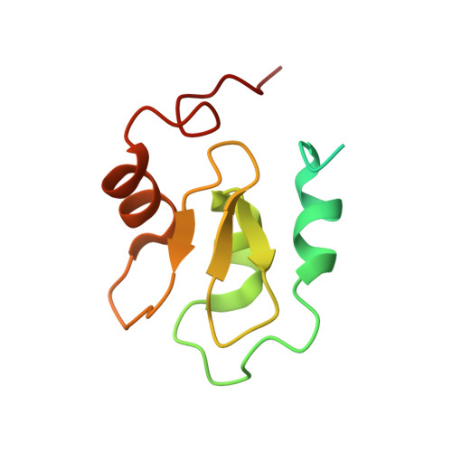
2QRA

Crystal structure of XIAP BIR1 domain (P21 form)


Experimental Data Snapshot

  • Method: X-RAY DIFFRACTION
  • Resolution: 2.50 Å
  • R-Value Free: 0.253 
  • R-Value Work: 0.196 

Starting Model: experimental
View more details

wwPDB Validation   3D Report Full Report


This is version 1.4 of the entry. See complete history


Literature

Crystal structure of the BIR1 domain of XIAP in two crystal forms

Lin, S.C.Huang, Y.Lo, Y.C.Lu, M.Wu, H.

(2007) J Mol Biol 372: 847-854

  • DOI: https://doi.org/10.1016/j.jmb.2007.07.019
  • Primary Citation of Related Structures:  
    2QRA

  • PubMed Abstract: 

    X-linked inhibitor of apoptosis (XIAP) is a potent negative regulator of apoptosis. It also plays a role in BMP signaling, TGF-beta signaling, and copper homeostasis. Previous structural studies have shown that the baculoviral IAP repeat (BIR2 and BIR3) domains of XIAP interact with the IAP-binding-motifs (IBM) in several apoptosis proteins such as Smac and caspase-9 via the conserved IBM-binding groove. Here, we report the crystal structure in two crystal forms of the BIR1 domain of XIAP, which does not possess this IBM-binding groove and cannot interact with Smac or caspase-9. Instead, the BIR1 domain forms a conserved dimer through the region corresponding to the IBM-binding groove. Structural and sequence analyses suggest that this dimerization of BIR1 in XIAP may be conserved in other IAP family members such as cIAP1 and cIAP2 and may be important for the action of XIAP in TGF-beta and BMP signaling and the action of cIAP1 and cIAP2 in TNF receptor signaling.


  • Organizational Affiliation

    Department of Biochemistry, Weill Medical College of Cornell University, New York, NY 10021, USA.


Macromolecules
Find similar proteins by:  (by identity cutoff)  |  3D Structure
Entity ID: 1
MoleculeChains Sequence LengthOrganismDetailsImage
Baculoviral IAP repeat-containing protein 4A [auth D],
B [auth C],
C [auth B],
D [auth A]
111Homo sapiensMutation(s): 0 
Gene Names: BIRC4API3IAP3XIAP
EC: 2.3.2.27
UniProt & NIH Common Fund Data Resources
Find proteins for P98170 (Homo sapiens)
Explore P98170 
Go to UniProtKB:  P98170
PHAROS:  P98170
GTEx:  ENSG00000101966 
Entity Groups  
Sequence Clusters30% Identity50% Identity70% Identity90% Identity95% Identity100% Identity
UniProt GroupP98170
Sequence Annotations
Expand
  • Reference Sequence
Small Molecules
Ligands 2 Unique
IDChains Name / Formula / InChI Key2D Diagram3D Interactions
ZN
Query on ZN

Download Ideal Coordinates CCD File 
E [auth D],
G [auth C],
K [auth B],
L [auth A]
ZINC ION
Zn
PTFCDOFLOPIGGS-UHFFFAOYSA-N
EOH
Query on EOH

Download Ideal Coordinates CCD File 
F [auth D]
H [auth C]
I [auth C]
J [auth C]
M [auth A]
F [auth D],
H [auth C],
I [auth C],
J [auth C],
M [auth A],
N [auth A]
ETHANOL
C2 H6 O
LFQSCWFLJHTTHZ-UHFFFAOYSA-N
Experimental Data & Validation

Experimental Data

  • Method: X-RAY DIFFRACTION
  • Resolution: 2.50 Å
  • R-Value Free: 0.253 
  • R-Value Work: 0.196 
  • Space Group: P 1 21 1
Unit Cell:
Length ( Å )Angle ( ˚ )
a = 36.563α = 90
b = 72.983β = 95.69
c = 68.904γ = 90
Software Package:
Software NamePurpose
DENZOdata reduction
SCALEPACKdata scaling
CNSrefinement
PDB_EXTRACTdata extraction
ADSCdata collection
CNSphasing

Structure Validation

View Full Validation Report


Entry History 

Deposition Data

  • Released Date: 2007-09-18 
  • Deposition Author(s): Lin, S.-C.

Revision History  (Full details and data files)

  • Version 1.0: 2007-09-18
    Type: Initial release
  • Version 1.1: 2011-07-13
    Changes: Version format compliance
  • Version 1.2: 2017-10-25
    Changes: Refinement description
  • Version 1.3: 2023-08-30
    Changes: Data collection, Database references, Derived calculations, Refinement description
  • Version 1.4: 2023-11-29
    Changes: Database references