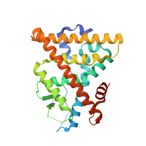
2QSE

Crystal Structure of the Estrogen Receptor Alpha Ligand Binding Domain complexed with Burned Meat Compound 4-OH-PhIP


Experimental Data Snapshot

  • Method: X-RAY DIFFRACTION
  • Resolution: 1.85 Å
  • R-Value Free: 0.225 
  • R-Value Work: 0.169 
  • R-Value Observed: 0.171 

Starting Model: experimental
View more details

wwPDB Validation   3D Report Full Report


Ligand Structure Quality Assessment 


This is version 1.4 of the entry. See complete history


Literature

NFkappaB selectivity of estrogen receptor ligands revealed by comparative crystallographic analyses

Nettles, K.W.Bruning, J.B.Gil, G.Nowak, J.Sharma, S.K.Hahm, J.B.Kulp, K.Hochberg, R.B.Zhou, H.Katzenellenbogen, J.A.Katzenellenbogen, B.S.Kim, Y.Joachmiak, A.Greene, G.L.

(2008) Nat Chem Biol 4: 241-247

  • DOI: https://doi.org/10.1038/nchembio.76
  • Primary Citation of Related Structures:  
    2B23, 2QA6, 2QA8, 2QAB, 2QGT, 2QGW, 2QH6, 2QR9, 2QSE, 2QXM

  • PubMed Abstract: 

    Our understanding of how steroid hormones regulate physiological functions has been significantly advanced by structural biology approaches. However, progress has been hampered by misfolding of the ligand binding domains in heterologous expression systems and by conformational flexibility that interferes with crystallization. Here, we show that protein folding problems that are common to steroid hormone receptors are circumvented by mutations that stabilize well-characterized conformations of the receptor. We use this approach to present the structure of an apo steroid receptor that reveals a ligand-accessible channel allowing soaking of preformed crystals. Furthermore, crystallization of different pharmacological classes of compounds allowed us to define the structural basis of NFkappaB-selective signaling through the estrogen receptor, thus revealing a unique conformation of the receptor that allows selective suppression of inflammatory gene expression. The ability to crystallize many receptor-ligand complexes with distinct pharmacophores allows one to define structural features of signaling specificity that would not be apparent in a single structure.


  • Organizational Affiliation

    Department of Cancer Biology, The Scripps Research Institute, 5353 Parkside Drive, Jupiter, Florida 33458, USA. knettles@scripps.edu


Macromolecules
Find similar proteins by:  (by identity cutoff)  |  3D Structure
Entity ID: 1
MoleculeChains Sequence LengthOrganismDetailsImage
Estrogen receptor
A, B
258Homo sapiensMutation(s): 1 
Gene Names: ESR1ESRNR3A1
UniProt & NIH Common Fund Data Resources
Find proteins for P03372 (Homo sapiens)
Explore P03372 
Go to UniProtKB:  P03372
PHAROS:  P03372
GTEx:  ENSG00000091831 
Entity Groups  
Sequence Clusters30% Identity50% Identity70% Identity90% Identity95% Identity100% Identity
UniProt GroupP03372
Sequence Annotations
Expand
  • Reference Sequence

Find similar proteins by:  Sequence   |   3D Structure  

Entity ID: 2
MoleculeChains Sequence LengthOrganismDetailsImage
Nuclear receptor coactivator 2
C, D
13Mus musculusMutation(s): 0 
UniProt & NIH Common Fund Data Resources
Find proteins for Q61026 (Mus musculus)
Explore Q61026 
Go to UniProtKB:  Q61026
IMPC:  MGI:1276533
Entity Groups  
Sequence Clusters30% Identity50% Identity70% Identity90% Identity95% Identity100% Identity
UniProt GroupQ61026
Sequence Annotations
Expand
  • Reference Sequence
Small Molecules
Ligands 1 Unique
IDChains Name / Formula / InChI Key2D Diagram3D Interactions
1HP
Query on 1HP

Download Ideal Coordinates CCD File 
E [auth A],
F [auth B]
4-(2-amino-1-methyl-1H-imidazo[4,5-b]pyridin-6-yl)phenol
C13 H12 N4 O
UGJXOCBVCWTJFP-UHFFFAOYSA-N
Experimental Data & Validation

Experimental Data

  • Method: X-RAY DIFFRACTION
  • Resolution: 1.85 Å
  • R-Value Free: 0.225 
  • R-Value Work: 0.169 
  • R-Value Observed: 0.171 
  • Space Group: P 1 21 1
Unit Cell:
Length ( Å )Angle ( ˚ )
a = 56.081α = 90
b = 84.044β = 108.85
c = 58.668γ = 90
Software Package:
Software NamePurpose
DENZOdata reduction
SCALEPACKdata scaling
PHASERphasing
REFMACrefinement
PDB_EXTRACTdata extraction
HKL-2000data collection
HKL-2000data reduction

Structure Validation

View Full Validation Report


Ligand Structure Quality Assessment 


Entry History 

Deposition Data

Revision History  (Full details and data files)

  • Version 1.0: 2008-03-18
    Type: Initial release
  • Version 1.1: 2011-07-13
    Changes: Advisory, Version format compliance
  • Version 1.2: 2017-10-25
    Changes: Refinement description, Source and taxonomy
  • Version 1.3: 2021-10-20
    Changes: Database references, Derived calculations
  • Version 1.4: 2023-08-30
    Changes: Data collection, Refinement description