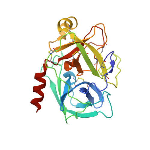
2QXH

Crystal Structure of Human Kallikrein 7 in Complex with Suc-Ala-Ala-Pro-Phe-chloromethylketone


Experimental Data Snapshot

  • Method: X-RAY DIFFRACTION
  • Resolution: 2.00 Å
  • R-Value Free: 0.233 
  • R-Value Work: 0.180 
  • R-Value Observed: 0.180 

Starting Model: experimental
View more details

wwPDB Validation   3D Report Full Report


Ligand Structure Quality Assessment 


This is version 1.3 of the entry. See complete history


Literature

Chymotryptic specificity determinants in the 1.0 A structure of the zinc-inhibited human tissue kallikrein 7.

Debela, M.Hess, P.Magdolen, V.Schechter, N.M.Steiner, T.Huber, R.Bode, W.Goettig, P.

(2007) Proc Natl Acad Sci U S A 104: 16086-16091

  • DOI: https://doi.org/10.1073/pnas.0707811104
  • Primary Citation of Related Structures:  
    2QXG, 2QXH, 2QXI, 2QXJ

  • PubMed Abstract: 

    hK7 or human stratum corneum chymotryptic enzyme belongs to the human tissue kallikrein (hKs) serine proteinase family and is strongly expressed in the upper layers of the epidermis. It participates in skin desquamation but is also implicated in diverse skin diseases and is a potential biomarker of ovarian cancer. We have solved x-ray structures of recombinant active hK7 at medium and atomic resolution in the presence of the inhibitors succinyl-Ala-Ala-Pro-Phe-chloromethyl ketone and Ala-Ala-Phe-chloromethyl ketone. The most distinguishing features of hK7 are the short 70-80 loop and the unique S1 pocket, which prefers P1 Tyr residues, as shown by kinetic data. Similar to several other kallikreins, the enzyme activity is inhibited by Zn(2+) and Cu(2+) at low micromolar concentrations. Biochemical analyses of the mutants H99A and H41F confirm that only the metal-binding site at His(99) close to the catalytic triad accounts for the noncompetitive Zn(2+) inhibition type. Additionally, hK7 exhibits large positively charged surface patches, representing putative exosites for prime side substrate recognition.


  • Organizational Affiliation

    Max-Planck-Institut für Biochemie, Proteinase Research Group, Am Klopferspitz 18, 82152 Martinsried, Germany.


Macromolecules
Find similar proteins by:  (by identity cutoff)  |  3D Structure
Entity ID: 1
MoleculeChains Sequence LengthOrganismDetailsImage
Kallikrein-7224Homo sapiensMutation(s): 0 
Gene Names: KLK7PRSS6SCCE
EC: 3.4.21.117
UniProt & NIH Common Fund Data Resources
Find proteins for P49862 (Homo sapiens)
Explore P49862 
Go to UniProtKB:  P49862
PHAROS:  P49862
GTEx:  ENSG00000169035 
Entity Groups  
Sequence Clusters30% Identity50% Identity70% Identity90% Identity95% Identity100% Identity
UniProt GroupP49862
Sequence Annotations
Expand
  • Reference Sequence
Small Molecules
Ligands 1 Unique
IDChains Name / Formula / InChI Key2D Diagram3D Interactions
K7J
Query on K7J

Download Ideal Coordinates CCD File 
B [auth A]N-(3-carboxypropanoyl)-L-alanyl-L-alanyl-N-[(2S,3S)-4-chloro-3-hydroxy-1-phenylbutan-2-yl]-L-prolinamide
C25 H35 Cl N4 O7
NDDWTTUSJLUGKF-CZKCSJLSSA-N
Experimental Data & Validation

Experimental Data

  • Method: X-RAY DIFFRACTION
  • Resolution: 2.00 Å
  • R-Value Free: 0.233 
  • R-Value Work: 0.180 
  • R-Value Observed: 0.180 
  • Space Group: P 1 21 1
Unit Cell:
Length ( Å )Angle ( ˚ )
a = 38.266α = 90
b = 56.964β = 101.37
c = 45.54γ = 90
Software Package:
Software NamePurpose
CNSrefinement
DENZOdata reduction
SCALEPACKdata scaling
PHASERphasing

Structure Validation

View Full Validation Report


Ligand Structure Quality Assessment 


Entry History 

Deposition Data

Revision History  (Full details and data files)

  • Version 1.0: 2008-01-08
    Type: Initial release
  • Version 1.1: 2011-07-13
    Changes: Version format compliance
  • Version 1.2: 2023-08-30
    Changes: Data collection, Database references, Derived calculations, Refinement description
  • Version 1.3: 2024-11-20
    Changes: Structure summary