2R5T

Crystal Structure of Inactive Serum and Glucocorticoid- Regulated Kinase 1 in Complex with AMP-PNP


Experimental Data Snapshot

  • Method: X-RAY DIFFRACTION
  • Resolution: 1.90 Å
  • R-Value Free: 0.215 
  • R-Value Work: 0.191 
  • R-Value Observed: 0.193 

wwPDB Validation   3D Report Full Report


Ligand Structure Quality Assessment 


This is version 1.4 of the entry. See complete history


Literature

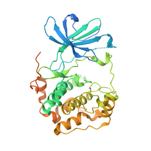
Crystal structure of the kinase domain of serum and glucocorticoid-regulated kinase 1 in complex with AMP PNP.

Zhao, B.Lehr, R.Smallwood, A.M.Ho, T.F.Maley, K.Randall, T.Head, M.S.Koretke, K.K.Schnackenberg, C.G.

(2007) Protein Sci 16: 2761-2769

  • DOI: https://doi.org/10.1110/ps.073161707
  • Primary Citation of Related Structures:  
    2R5T

  • PubMed Abstract: 
  • Serum and glucocorticoid-regulated kinase 1 (SGK1) is a serine/threonine protein kinase of the AGC family which participates in the control of epithelial ion transport and is implicated in proliferation and apoptosis. We report here the 1.9 A crystal structure of the catalytic domain of inactive human SGK1 in complex with AMP-PNP ...

    Serum and glucocorticoid-regulated kinase 1 (SGK1) is a serine/threonine protein kinase of the AGC family which participates in the control of epithelial ion transport and is implicated in proliferation and apoptosis. We report here the 1.9 A crystal structure of the catalytic domain of inactive human SGK1 in complex with AMP-PNP. SGK1 exists as a dimer formed by two intermolecular disulfide bonds between Cys258 in the activation loop and Cys193. Although most of the SGK1 structure closely resembles the common protein kinase fold, the structure around the active site is unique when compared to most protein kinases. The alphaC helix is not present in this inactive form of SGK1 crystal structure; instead, the segment corresponding to the C helix forms a beta-strand that is stabilized by the N-terminal segment of the activation loop through a short antiparallel beta-sheet. Since the differences from other kinases occur around the ATP binding site, this structure can provide valuable insight into the design of selective and highly potent ATP-competitive inhibitors of SGK1 kinase.


    Organizational Affiliation

    Department of Computational and Structural Chemistry, GlaxoSmithKline, King of Prussia, Pennsylvania 19406, USA. baoguang.zhao@gsk.com


Macromolecules
Find similar proteins by:  (by identity cutoff)  |  3D Structure
Entity ID: 1
MoleculeChainsSequence LengthOrganismDetailsImage
Serine/threonine-protein kinase Sgk1373Homo sapiensMutation(s): 4 
Gene Names: SGKSGK1
EC: 2.7.11.1
UniProt & NIH Common Fund Data Resources
Find proteins for O00141 (Homo sapiens)
Explore O00141 
Go to UniProtKB:  O00141
PHAROS:  O00141
Entity Groups  
Sequence Clusters30% Identity50% Identity70% Identity90% Identity95% Identity100% Identity
UniProt GroupO00141
Protein Feature View
Expand
  • Reference Sequence
Small Molecules
Ligands 3 Unique
IDChainsName / Formula / InChI Key2D Diagram3D Interactions
ANP
Query on ANP

Download Ideal Coordinates CCD File 
D [auth A]PHOSPHOAMINOPHOSPHONIC ACID-ADENYLATE ESTER
C10 H17 N6 O12 P3
PVKSNHVPLWYQGJ-KQYNXXCUSA-N
 Ligand Interaction
SO4
Query on SO4

Download Ideal Coordinates CCD File 
C [auth A]SULFATE ION
O4 S
QAOWNCQODCNURD-UHFFFAOYSA-L
 Ligand Interaction
MG
Query on MG

Download Ideal Coordinates CCD File 
B [auth A]MAGNESIUM ION
Mg
JLVVSXFLKOJNIY-UHFFFAOYSA-N
 Ligand Interaction
Experimental Data & Validation

Experimental Data

  • Method: X-RAY DIFFRACTION
  • Resolution: 1.90 Å
  • R-Value Free: 0.215 
  • R-Value Work: 0.191 
  • R-Value Observed: 0.193 
  • Space Group: P 65 2 2
Unit Cell:
Length ( Å )Angle ( ˚ )
a = 97.873α = 90
b = 97.873β = 90
c = 147.848γ = 120
Software Package:
Software NamePurpose
REFMACrefinement
SCALEPACKdata scaling
CNSrefinement
DENZOdata reduction
PDB_EXTRACTdata extraction
HKL-2000data collection
HKL-2000data reduction
CNSphasing

Structure Validation

View Full Validation Report


Ligand Structure Quality Assessment 


Entry History 

Deposition Data

Revision History  (Full details and data files)

  • Version 1.0: 2008-09-09
    Type: Initial release
  • Version 1.1: 2011-07-13
    Changes: Version format compliance
  • Version 1.2: 2017-10-25
    Changes: Refinement description
  • Version 1.3: 2019-07-24
    Changes: Data collection, Derived calculations, Refinement description
  • Version 1.4: 2021-10-20
    Changes: Database references, Derived calculations