Structural Characterization of a New Binding Motif and a Novel Binding Mode in Group 2 WW Domains
Ramirez-Espain, X., Ruiz, L., Martin-Malpartida, P., Oschkinat, H., Macias, M.J.(2007) J Mol Biol 373: 1255-1268
- PubMed: 17915251
- DOI: https://doi.org/10.1016/j.jmb.2007.08.052
- Primary Citation of Related Structures:
2JUP, 2RLY, 2RM0 - PubMed Abstract:
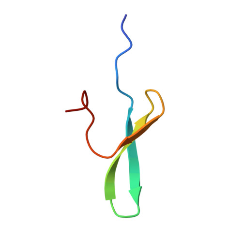
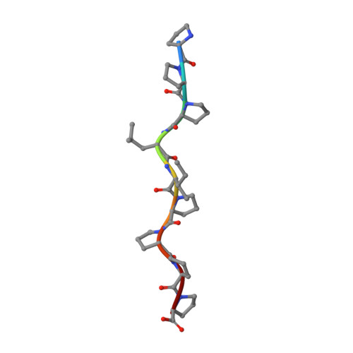
Formin homology 1 (FH1), is a long proline-rich region of formins, shown to bind to five WW containing proteins named formin binding proteins (FBPs). FH1 has several potential binding regions but only the PPLPx motif and its interaction with FBP11WW1 has been characterized structurally. To detect whether additional motifs exist in FH1, we synthesized five peptides and investigated their interaction with FBP28WW2, FBP11WW1 and FBP11WW2 domains. Peptides of sequence PTPPPLPP (positive control), PPPLIPPPP and PPLIPPPP (new motifs) interact with the domains with micromolar affinity. We observed that FBP28WW2 and FBP11WW2 behave differently from FBP11WW1 in terms of motif selection and affinity, since they prefer a doubly interrupted proline stretch of sequence PPLIPP. We determined the NMR structure of three complexes involving the FBP28WW2 domain and the three ligands. Depending on the peptide under study, the domain interacts with two proline residues accommodated in either the XP or the XP2 groove. This difference represents a one-turn displacement of the domain along the ligand sequence. To understand what drives this behavior, we performed further structural studies with the FBP11WW1 and a mutant of FBP28WW2 mimicking the XP2 groove of FBP11WW1. Our observations suggest that the nature of the XP2 groove and the balance of flexibility/rigidity around loop 1 of the domain contribute to the selection of the final ligand positioning in fully independent domains. Additionally, we analyzed the binding of a double WW domain region, FBP11WW1-2, to a long stretch of FH1 using fluorescence spectroscopy and NMR titrations. With this we show that the presence of two consecutive WW domains may also influence the selection of the binding mode, particularly if both domains can interact with consecutive motifs in the ligand. Our results represent the first observation of protein-ligand recognition where a pair of WW and two consecutive motifs in a ligand participate simultaneously.
Organizational Affiliation:
Institute for Research in Biomedicine-Protein NMR group, and the Institució catalana de recerca i estudis avançats ICREA, Barcelona Science Park, Josep Samitier 1-5, E-08028 Barcelona, Spain.


