2V55 | pdb_00002v55

Mechanism of multi-site phosphorylation from a ROCK-I:RhoE complex structure


Experimental Data Snapshot

  • Method: X-RAY DIFFRACTION
  • Resolution: 3.71 Å
  • R-Value Free: 
    0.269 (Depositor), 0.264 (DCC) 
  • R-Value Work: 
    0.238 (Depositor), 0.229 (DCC) 
  • R-Value Observed: 
    0.238 (Depositor) 

Starting Models: experimental
View more details

wwPDB Validation 3D Report Full Report

Validation slider image for 2V55

Ligand Structure Quality Assessment 


This is version 1.3 of the entry. See complete history

Literature

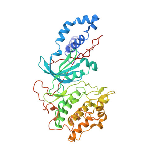
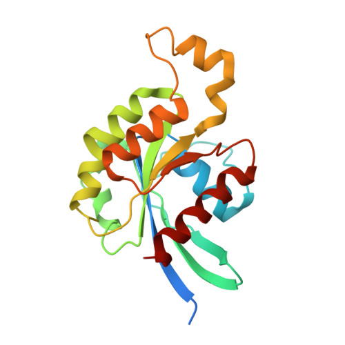
Mechanism of Multi-Site Phosphorylation from a Rock-I:Rhoe Complex Structure.

Komander, D.Garg, R.Wan, P.T.C.Ridley, A.J.Barford, D.

(2008) EMBO J 27: 3175

  • DOI: https://doi.org/10.1038/emboj.2008.226
  • Primary Citation Related Structures: 
    2V55

  • PubMed Abstract: 

    The ROCK-I serine/threonine protein kinase mediates the effects of RhoA to promote the formation of actin stress fibres and integrin-based focal adhesions. ROCK-I phosphorylates the unconventional G-protein RhoE on multiple N- and C-terminal sites. These phosphorylation events stabilise RhoE, which functions to antagonise RhoA-induced stress fibre assembly. Here, we provide a molecular explanation for multi-site phosphorylation of RhoE from the crystal structure of RhoE in complex with the ROCK-I kinase domain. RhoE interacts with the C-lobe alphaG helix of ROCK-I by means of a novel binding site remote from its effector region, positioning its N and C termini proximal to the ROCK-I catalytic site. Disruption of the ROCK-I:RhoE interface abolishes RhoE phosphorylation, but has no effect on the ability of RhoE to disassemble stress fibres. In contrast, mutation of the RhoE effector region attenuates RhoE-mediated disruption of the actin cytoskeleton, indicating that RhoE exerts its inhibitory effects on ROCK-I through protein(s) binding to its effector region. We propose that ROCK-I phosphorylation of RhoE forms part of a feedback loop to regulate RhoA signalling.


  • Organizational Affiliation
    • Section of Structural Biology, The Institute of Cancer Research, Chester Beatty Laboratories, London, UK.

Macromolecule Content 

  • Total Structure Weight: 141.15 kDa 
  • Atom Count: 9,188 
  • Modeled Residue Count: 1,152 
  • Deposited Residue Count: 1,212 
  • Unique protein chains: 2

Macromolecules

Find similar proteins by:|  3D Structure
Entity ID: 1
MoleculeChains  Sequence LengthOrganismDetailsImage
RHO-ASSOCIATED PROTEIN KINASE 1
A, C
406Homo sapiensMutation(s): 0 
EC: 2.7.11.1 (PDB Primary Data), 2.7.11.39 (UniProt)
UniProt & NIH Common Fund Data Resources
Find proteins for Q13464 (Homo sapiens)
Explore Q13464 
Go to UniProtKB:  Q13464
PHAROS:  Q13464
GTEx:  ENSG00000067900 
Entity Groups
Sequence Clusters30% Identity50% Identity70% Identity90% Identity95% Identity100% Identity
UniProt GroupQ13464
Sequence Annotations
Expand
Reference Sequence
Find similar proteins by:|  3D Structure
Entity ID: 2
MoleculeChains  Sequence LengthOrganismDetailsImage
RHO-RELATED GTP-BINDING PROTEIN RHOE
B, D
200Homo sapiensMutation(s): 0 
UniProt & NIH Common Fund Data Resources
Find proteins for P61587 (Homo sapiens)
Explore P61587 
Go to UniProtKB:  P61587
PHAROS:  P61587
GTEx:  ENSG00000115963 
Entity Groups
Sequence Clusters30% Identity50% Identity70% Identity90% Identity95% Identity100% Identity
UniProt GroupP61587
Sequence Annotations
Expand
Reference Sequence

Small Molecules

Ligands 4 Unique
IDChains Name / Formula / InChI Key2D Diagram3D Interactions
GTP

Query on GTP


Download:Ideal Coordinates CCD File
G [auth B],
K [auth D]
GUANOSINE-5'-TRIPHOSPHATE
C10 H16 N5 O14 P3
XKMLYUALXHKNFT-UUOKFMHZSA-N
ANP

Query on ANP


Download:Ideal Coordinates CCD File
E [auth A],
I [auth C]
PHOSPHOAMINOPHOSPHONIC ACID-ADENYLATE ESTER
C10 H17 N6 O12 P3
PVKSNHVPLWYQGJ-KQYNXXCUSA-N
SO4

Query on SO4


Download:Ideal Coordinates CCD File
M [auth D]SULFATE ION
O4 S
QAOWNCQODCNURD-UHFFFAOYSA-L
MG

Query on MG


Download:Ideal Coordinates CCD File
F [auth A],
H [auth B],
J [auth C],
L [auth D]
MAGNESIUM ION
Mg
JLVVSXFLKOJNIY-UHFFFAOYSA-N

Experimental Data & Validation

Experimental Data

  • Method: X-RAY DIFFRACTION
  • Resolution: 3.71 Å
  • R-Value Free:  0.269 (Depositor), 0.264 (DCC) 
  • R-Value Work:  0.238 (Depositor), 0.229 (DCC) 
  • R-Value Observed: 0.238 (Depositor) 
Space Group: P 65 2 2
Unit Cell:
Length ( Å )Angle ( ˚ )
a = 152.477α = 90
b = 152.477β = 90
c = 531.274γ = 120
Software Package:
Software NamePurpose
PHENIXrefinement
MOSFLMdata reduction
SCALAdata scaling
PHASERphasing

Structure Validation

View Full Validation Report


Ligand Structure Quality Assessment 


Entry History 

Deposition Data

Revision History  (Full details and data files)

  • Version 1.0: 2008-11-04
    Type: Initial release
  • Version 1.1: 2011-05-08
    Changes: Version format compliance
  • Version 1.2: 2011-07-13
    Changes: Version format compliance
  • Version 1.3: 2023-12-13
    Changes: Data collection, Database references, Derived calculations, Other, Refinement description