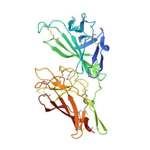
2V5N

STRUCTURE OF HUMAN IGF2R DOMAINS 11-12


Experimental Data Snapshot

  • Method: X-RAY DIFFRACTION
  • Resolution: 3.20 Å
  • R-Value Free: 0.317 
  • R-Value Work: 0.253 
  • R-Value Observed: 0.259 

wwPDB Validation   3D Report Full Report


Ligand Structure Quality Assessment 


This is version 1.4 of the entry. See complete history


Literature

Structure and Functional Analysis of the Igf-II/Igf2R Interaction

Brown, J.Delaine, C.Zaccheo, O.J.Siebold, C.Gilbert, R.J.Van Boxel, G.Denley, A.Wallace, J.C.Hassan, A.B.Forbes, B.E.Jones, E.Y.

(2008) EMBO J 27: 265

  • DOI: https://doi.org/10.1038/sj.emboj.7601938
  • Primary Citation of Related Structures:  
    2V5N, 2V5O, 2V5P

  • PubMed Abstract: 

    Embryonic development and normal growth require exquisite control of insulin-like growth factors (IGFs). In mammals the extracellular region of the cation-independent mannose-6-phosphate receptor has gained an IGF-II-binding function and is termed type II IGF receptor (IGF2R). IGF2R sequesters IGF-II; imbalances occur in cancers and IGF2R is implicated in tumour suppression. We report crystal structures of IGF2R domains 11-12, 11-12-13-14 and domains 11-12-13/IGF-II complex. A distinctive juxtaposition of these domains provides the IGF-II-binding unit, with domain 11 directly interacting with IGF-II and domain 13 modulating binding site flexibility. Our complex shows that Phe19 and Leu53 of IGF-II lock into a hydrophobic pocket unique to domain 11 of mammalian IGF2Rs. Mutagenesis analyses confirm this IGF-II 'binding-hotspot', revealing that IGF-binding proteins and IGF2R have converged on the same high-affinity site.


  • Organizational Affiliation

    Cancer Research UK Receptor Structure Research Group, Division of Structural Biology, Wellcome Trust Centre for Human Genetics, University of Oxford, Headington, Oxford, UK.


Macromolecules
Find similar proteins by:  (by identity cutoff)  |  3D Structure
Entity ID: 1
MoleculeChains Sequence LengthOrganismDetailsImage
CATION-INDEPENDENT MANNOSE-6-PHOSPHATE RECEPTOR299Homo sapiensMutation(s): 0 
UniProt & NIH Common Fund Data Resources
Find proteins for P11717 (Homo sapiens)
Explore P11717 
Go to UniProtKB:  P11717
PHAROS:  P11717
GTEx:  ENSG00000197081 
Entity Groups  
Sequence Clusters30% Identity50% Identity70% Identity90% Identity95% Identity100% Identity
UniProt GroupP11717
Glycosylation
Glycosylation Sites: 2Go to GlyGen: P11717-1
Sequence Annotations
Expand
  • Reference Sequence
Small Molecules
Modified Residues  1 Unique
IDChains TypeFormula2D DiagramParent
MSE
Query on MSE
A
L-PEPTIDE LINKINGC5 H11 N O2 SeMET
Experimental Data & Validation

Experimental Data

  • Method: X-RAY DIFFRACTION
  • Resolution: 3.20 Å
  • R-Value Free: 0.317 
  • R-Value Work: 0.253 
  • R-Value Observed: 0.259 
  • Space Group: P 61 2 2
Unit Cell:
Length ( Å )Angle ( ˚ )
a = 64.793α = 90
b = 64.793β = 90
c = 269.569γ = 120
Software Package:
Software NamePurpose
REFMACrefinement
XDSdata reduction
XDSdata scaling
HKL2Mapphasing

Structure Validation

View Full Validation Report


Ligand Structure Quality Assessment 


Entry History 

Deposition Data

Revision History  (Full details and data files)

  • Version 1.0: 2007-12-11
    Type: Initial release
  • Version 1.1: 2011-07-13
    Changes: Advisory, Version format compliance
  • Version 1.2: 2018-02-28
    Changes: Refinement description, Source and taxonomy
  • Version 1.3: 2020-07-29
    Type: Remediation
    Reason: Carbohydrate remediation
    Changes: Data collection, Derived calculations, Other, Structure summary
  • Version 1.4: 2024-11-20
    Changes: Data collection, Database references, Structure summary