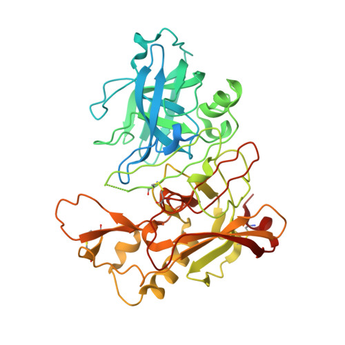
2VA5

X-ray crystal structure of beta secretase complexed with compound 8c


Experimental Data Snapshot

  • Method: X-RAY DIFFRACTION
  • Resolution: 2.75 Å
  • R-Value Free: 0.319 
  • R-Value Work: 0.228 
  • R-Value Observed: 0.232 

Starting Model: experimental
View more details

wwPDB Validation   3D Report Full Report


Ligand Structure Quality Assessment 


This is version 1.4 of the entry. See complete history


Literature

Application of Fragment-Based Lead Generation to the Discovery of Novel, Cyclic Amidine Beta-Secretase Inhibitors with Nanomolar Potency, Cellular Activity, and High Ligand Efficiency.

Edwards, P.D.Albert, J.S.Sylvester, M.Aharony, D.Andisik, D.Callaghan, O.Campbell, J.B.Carr, R.A.Chessari, G.Congreve, M.Frederickson, M.Folmer, R.H.Geschwindner, S.Koether, G.Kolmodin, K.Krumrine, J.Mauger, R.C.Murray, C.W.Olsson, L.L.Patel, S.Spear, N.Tian, G.

(2007) J Med Chem 50: 5912

  • DOI: https://doi.org/10.1021/jm070829p
  • Primary Citation of Related Structures:  
    2VA5, 2VA6, 2VA7

  • PubMed Abstract: 

    Fragment-based lead generation has led to the discovery of a novel series of cyclic amidine-based inhibitors of beta-secretase (BACE-1). Initial fragment hits with an isocytosine core having millimolar potency were identified via NMR affinity screening. Structure-guided evolution of these fragments using X-ray crystallography together with potency determination using surface plasmon resonance and functional enzyme inhibition assays afforded micromolar inhibitors. Similarity searching around the isocytosine core led to the identification of a related series of inhibitors, the dihydroisocytosines. By leveraging the knowledge of the ligand-BACE-1 recognition features generated from the isocytosines, the dihydroisocytosines were efficiently optimized to submicromolar potency. Compound 29, with an IC50 of 80 nM, a ligand efficiency of 0.37, and cellular activity of 470 nM, emerged as the lead structure for future optimization.


  • Organizational Affiliation

    CNS Discovery Research, AstraZeneca Pharmaceuticals LP, 1800 Concord Pike, Wilmington, DE 19850-5437, USA. philip.edwards@astrazeneca.com


Macromolecules
Find similar proteins by:  (by identity cutoff)  |  3D Structure
Entity ID: 1
MoleculeChains Sequence LengthOrganismDetailsImage
BETA-SECRETASE 1 .455Homo sapiensMutation(s): 2 
EC: 3.4.23.46
UniProt & NIH Common Fund Data Resources
Find proteins for P56817 (Homo sapiens)
Explore P56817 
Go to UniProtKB:  P56817
PHAROS:  P56817
GTEx:  ENSG00000186318 
Entity Groups  
Sequence Clusters30% Identity50% Identity70% Identity90% Identity95% Identity100% Identity
UniProt GroupP56817
Sequence Annotations
Expand
  • Reference Sequence
Small Molecules
Binding Affinity Annotations 
IDSourceBinding Affinity
C8C PDBBind:  2VA5 IC50: 8.60e+4 (nM) from 1 assay(s)
BindingDB:  2VA5 IC50: min: 8.60e+4, max: 1.30e+5 (nM) from 2 assay(s)
Experimental Data & Validation

Experimental Data

  • Method: X-RAY DIFFRACTION
  • Resolution: 2.75 Å
  • R-Value Free: 0.319 
  • R-Value Work: 0.228 
  • R-Value Observed: 0.232 
  • Space Group: P 61 2 2
Unit Cell:
Length ( Å )Angle ( ˚ )
a = 102.138α = 90
b = 102.138β = 90
c = 168.258γ = 120
Software Package:
Software NamePurpose
REFMACrefinement
MOSFLMdata reduction
SCALAdata scaling
CSearchphasing

Structure Validation

View Full Validation Report


Ligand Structure Quality Assessment 


Entry History 

Revision History  (Full details and data files)

  • Version 1.0: 2007-11-13
    Type: Initial release
  • Version 1.1: 2011-05-08
    Changes: Version format compliance
  • Version 1.2: 2011-07-13
    Changes: Version format compliance
  • Version 1.3: 2023-12-13
    Changes: Data collection, Database references, Other, Refinement description
  • Version 1.4: 2024-11-06
    Changes: Refinement description, Structure summary