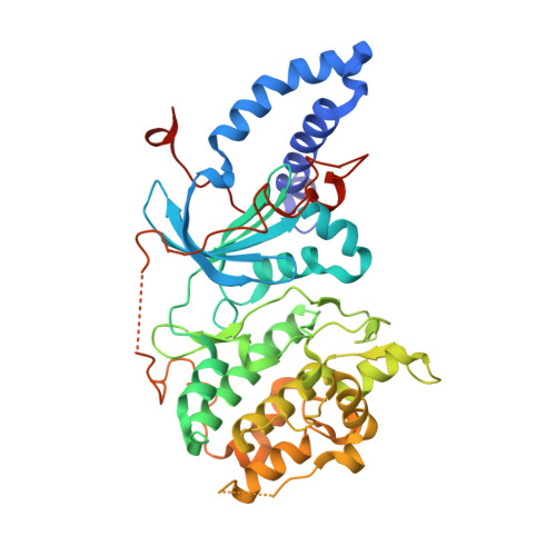
2VD5

Structure of Human Myotonic Dystrophy Protein Kinase in Complex with the Bisindoylmaleide inhibitor BIM VIII


Experimental Data Snapshot

  • Method: X-RAY DIFFRACTION
  • Resolution: 2.80 Å
  • R-Value Free: 0.244 
  • R-Value Work: 0.196 
  • R-Value Observed: 0.198 

Starting Model: experimental
View more details

wwPDB Validation   3D Report Full Report


Ligand Structure Quality Assessment 


This is version 1.2 of the entry. See complete history


Literature

Structure of Dystrophia Myotonica Protein Kinase.

Elkins, J.Amos, A.Niesen, F.Pike, A.C.W.Fedorov, O.Knapp, S.

(2009) Protein Sci 18: 782

  • DOI: https://doi.org/10.1002/pro.82
  • Primary Citation of Related Structures:  
    2VD5

  • PubMed Abstract: 

    Dystrophia myotonica protein kinase (DMPK) is a serine/threonine kinase composed of a kinase domain and a coiled-coil domain involved in the multimerization. The crystal structure of the kinase domain of DMPK bound to the inhibitor bisindolylmaleimide VIII (BIM-8) revealed a dimeric enzyme associated by a conserved dimerization domain. The affinity of dimerisation suggested that the kinase domain alone is insufficient for dimerisation in vivo and that the coiled-coil domains are required for stable dimer formation. The kinase domain is in an active conformation, with a fully-ordered and correctly positioned alphaC helix, and catalytic residues in a conformation competent for catalysis. The conserved hydrophobic motif at the C-terminal extension of the kinase domain is bound to the N-terminal lobe of the kinase domain, despite being unphosphorylated. Differences in the arrangement of the C-terminal extension compared to the closely related Rho-associated kinases include an altered PXXP motif, a different conformation and binding arrangement for the turn motif, and a different location for the conserved NFD motif. The BIM-8 inhibitor occupies the ATP site and has similar binding mode as observed in PDK1.


  • Organizational Affiliation

    Structural Genomics Consortium, Nuffield Department of Medicine, Oxford University, Old Road Campus Research Building, Oxford, OX3 7DQ, United Kingdom.


Macromolecules
Find similar proteins by:  (by identity cutoff)  |  3D Structure
Entity ID: 1
MoleculeChains Sequence LengthOrganismDetailsImage
DMPK PROTEIN
A, B
412Homo sapiensMutation(s): 0 
EC: 2.7.11.1
UniProt & NIH Common Fund Data Resources
Find proteins for Q09013 (Homo sapiens)
Explore Q09013 
Go to UniProtKB:  Q09013
PHAROS:  Q09013
GTEx:  ENSG00000104936 
Entity Groups  
Sequence Clusters30% Identity50% Identity70% Identity90% Identity95% Identity100% Identity
UniProt GroupQ09013
Sequence Annotations
Expand
  • Reference Sequence
Small Molecules
Ligands 1 Unique
IDChains Name / Formula / InChI Key2D Diagram3D Interactions
BI8
Query on BI8

Download Ideal Coordinates CCD File 
C [auth A],
D [auth B]
3-[1-(3-AMINOPROPYL)-1H-INDOL-3-YL]-4-(1-METHYL-1H-INDOL-3-YL)-1H-PYRROLE-2,5-DIONE
C24 H22 N4 O2
UQHKJRCFSLMWIA-UHFFFAOYSA-N
Experimental Data & Validation

Experimental Data

  • Method: X-RAY DIFFRACTION
  • Resolution: 2.80 Å
  • R-Value Free: 0.244 
  • R-Value Work: 0.196 
  • R-Value Observed: 0.198 
  • Space Group: P 32 2 1
Unit Cell:
Length ( Å )Angle ( ˚ )
a = 122.599α = 90
b = 122.599β = 90
c = 134.278γ = 120
Software Package:
Software NamePurpose
REFMACrefinement
MOSFLMdata reduction
SCALAdata scaling
PHASERphasing

Structure Validation

View Full Validation Report


Ligand Structure Quality Assessment 


Entry History 

Revision History  (Full details and data files)

  • Version 1.0: 2007-11-06
    Type: Initial release
  • Version 1.1: 2011-07-13
    Changes: Advisory, Version format compliance
  • Version 1.2: 2023-12-13
    Changes: Data collection, Database references, Derived calculations, Other, Refinement description