2VIF

Crystal structure of SOCS6 SH2 domain in complex with a c-KIT phosphopeptide


Experimental Data Snapshot

  • Method: X-RAY DIFFRACTION
  • Resolution: 1.45 Å
  • R-Value Free: 0.198 
  • R-Value Work: 0.139 
  • R-Value Observed: 0.142 

wwPDB Validation   3D Report Full Report


This is version 1.3 of the entry. See complete history


Literature

Structural Basis for C-Kit Inhibition by the Suppressor of Cytokine Signaling 6 (Socs6) Ubiquitin Ligase.

Zadjali, F.Pike, A.C.W.Vesterlund, M.Sun, J.Wu, C.Li, S.S.Ronnstrand, L.Knapp, S.Bullock, A.Flores-Morales, A.

(2011) J Biol Chem 286: 480

  • DOI: https://doi.org/10.1074/jbc.M110.173526
  • Primary Citation of Related Structures:  
    2VIF

  • PubMed Abstract: 

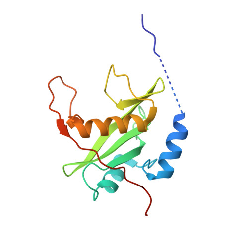
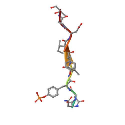
    The c-KIT receptor tyrosine kinase mediates the cellular response to stem cell factor (SCF). Whereas c-KIT activity is important for the proliferation of hematopoietic cells, melanocytes and germ cells, uncontrolled c-KIT activity contributes to the growth of diverse human tumors. Suppressor of cytokine signaling 6 (SOCS6) is a member of the SOCS family of E3 ubiquitin ligases that can interact with c-KIT and suppress c-KIT-dependent pathways. Here, we analyzed the molecular mechanisms that determine SOCS6 substrate recognition. Our results show that the SH2 domain of SOCS6 is essential for its interaction with c-KIT pY568. The 1.45-Å crystal structure of SOCS6 SH2 domain bound to the c-KIT substrate peptide (c-KIT residues 564-574) revealed a highly complementary and specific interface giving rise to a high affinity interaction (K(d) = 0.3 μm). Interestingly, the SH2 binding pocket extends to substrate residue position pY+6 and envelopes the c-KIT phosphopeptide with a large BG loop insertion that contributes significantly to substrate interaction. We demonstrate that SOCS6 has ubiquitin ligase activity toward c-KIT and regulates c-KIT protein turnover in cells. Our data support a role of SOCS6 as a feedback inhibitor of SCF-dependent signaling and provides molecular data to account for target specificity within the SOCS family of ubiquitin ligases.


  • Organizational Affiliation

    Department of Molecular Medicine and Surgery, Karolinska Institutet, SE-17177 Stockholm, Sweden.


Macromolecules
Find similar proteins by:  (by identity cutoff)  |  3D Structure
Entity ID: 1
MoleculeChains Sequence LengthOrganismDetailsImage
SUPPRESSOR OF CYTOKINE SIGNALLING 6141Homo sapiensMutation(s): 0 
UniProt & NIH Common Fund Data Resources
Find proteins for O14544 (Homo sapiens)
Explore O14544 
Go to UniProtKB:  O14544
PHAROS:  O14544
GTEx:  ENSG00000170677 
Entity Groups  
Sequence Clusters30% Identity50% Identity70% Identity90% Identity95% Identity100% Identity
UniProt GroupO14544
Sequence Annotations
Expand
  • Reference Sequence

Find similar proteins by:  Sequence   |   3D Structure  

Entity ID: 2
MoleculeChains Sequence LengthOrganismDetailsImage
MAST/STEM CELL GROWTH FACTOR RECEPTORB [auth P]11Homo sapiensMutation(s): 0 
UniProt & NIH Common Fund Data Resources
Find proteins for P10721 (Homo sapiens)
Explore P10721 
Go to UniProtKB:  P10721
PHAROS:  P10721
GTEx:  ENSG00000157404 
Entity Groups  
Sequence Clusters30% Identity50% Identity70% Identity90% Identity95% Identity100% Identity
UniProt GroupP10721
Sequence Annotations
Expand
  • Reference Sequence
Small Molecules
Ligands 1 Unique
IDChains Name / Formula / InChI Key2D Diagram3D Interactions
EDO
Query on EDO

Download Ideal Coordinates CCD File 
C [auth A]1,2-ETHANEDIOL
C2 H6 O2
LYCAIKOWRPUZTN-UHFFFAOYSA-N
Modified Residues  1 Unique
IDChains TypeFormula2D DiagramParent
PTR
Query on PTR
B [auth P]L-PEPTIDE LINKINGC9 H12 N O6 PTYR
Experimental Data & Validation

Experimental Data

  • Method: X-RAY DIFFRACTION
  • Resolution: 1.45 Å
  • R-Value Free: 0.198 
  • R-Value Work: 0.139 
  • R-Value Observed: 0.142 
  • Space Group: P 21 21 21
Unit Cell:
Length ( Å )Angle ( ˚ )
a = 30.377α = 90
b = 60.143β = 90
c = 71.367γ = 90
Software Package:
Software NamePurpose
REFMACrefinement
MOSFLMdata reduction
SCALAdata scaling
PHASERphasing

Structure Validation

View Full Validation Report


Entry History 

Deposition Data

Revision History  (Full details and data files)

  • Version 1.0: 2007-12-25
    Type: Initial release
  • Version 1.1: 2011-05-08
    Changes: Version format compliance
  • Version 1.2: 2011-07-13
    Changes: Version format compliance
  • Version 1.3: 2023-12-13
    Changes: Data collection, Database references, Derived calculations, Other, Refinement description