2VL9 | pdb_00002vl9

Oxidized form of human peroxiredoxin 5


Experimental Data Snapshot

  • Method: X-RAY DIFFRACTION
  • Resolution: 2.70 Å
  • R-Value Free: 
    0.239 (Depositor), 0.234 (DCC) 
  • R-Value Work: 
    0.184 (Depositor), 0.190 (DCC) 
  • R-Value Observed: 
    0.186 (Depositor) 

Starting Model: experimental
View more details

wwPDB Validation 3D Report Full Report

Validation slider image for 2VL9

This is version 1.5 of the entry. See complete history

Literature

The Crystal Structures of Oxidized Forms of Human Peroxiredoxin 5 with an Intramolecular Disulfide Bond Confirm the Proposed Enzymatic Mechanism for Atypical 2-Cys Peroxiredoxins.

Smeets, A.Marchand, C.Linard, D.Knoops, B.Declercq, J.P.

(2008) Arch Biochem Biophys 477: 98

  • DOI: https://doi.org/10.1016/j.abb.2008.04.036
  • Primary Citation Related Structures: 
    2VL2, 2VL3, 2VL9

  • PubMed Abstract: 

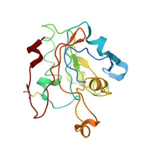
    Peroxiredoxin 5 (PRDX5) belongs to the PRDX superfamily of thiol-dependent peroxidases able to reduce hydrogen peroxide, alkyl hydroperoxides and peroxynitrite. PRDX5 is classified in the atypical 2-Cys subfamily of PRDXs. In this subfamily, the oxidized form of the enzyme is characterized by the presence of an intramolecular disulfide bridge between the peroxidatic and the resolving cysteine residues. We report here three crystal forms in which this intramolecular disulfide bond is indeed observed. The structures are characterized by the expected local unfolding of the peroxidatic loop, but also by the unfolding of the resolving loop. A new type of interface between PRDX molecules is described. The three crystal forms were not oxidized in the same way and the influence of the oxidizing conditions is discussed.


  • Organizational Affiliation
    • Unit of Structural Chemistry (CSTR), Université catholique de Louvain, 1, place Louis Pasteur, B-1348 Louvain-la-Neuve, Belgium.

Macromolecule Content 

  • Total Structure Weight: 73.24 kDa 
  • Atom Count: 4,817 
  • Modeled Residue Count: 644 
  • Deposited Residue Count: 692 
  • Unique protein chains: 1

Macromolecules

Find similar proteins by:|  3D Structure
Entity ID: 1
MoleculeChains  Sequence LengthOrganismDetailsImage
PEROXIREDOXIN-5
A, B, C, D
173Homo sapiensMutation(s): 1 
EC: 1.11.1.15 (PDB Primary Data), 1.11.1.24 (UniProt)
UniProt & NIH Common Fund Data Resources
Find proteins for P30044 (Homo sapiens)
Explore P30044 
Go to UniProtKB:  P30044
PHAROS:  P30044
GTEx:  ENSG00000126432 
Entity Groups
Sequence Clusters30% Identity50% Identity70% Identity90% Identity95% Identity100% Identity
UniProt GroupP30044
Sequence Annotations
Expand
Reference Sequence

Experimental Data & Validation

Experimental Data

  • Method: X-RAY DIFFRACTION
  • Resolution: 2.70 Å
  • R-Value Free:  0.239 (Depositor), 0.234 (DCC) 
  • R-Value Work:  0.184 (Depositor), 0.190 (DCC) 
  • R-Value Observed: 0.186 (Depositor) 
Space Group: P 31 2 1
Unit Cell:
Length ( Å )Angle ( ˚ )
a = 75.87α = 90
b = 75.87β = 90
c = 193.254γ = 120
Software Package:
Software NamePurpose
PHENIXrefinement
XDSdata reduction
XSCALEdata scaling
PHASERphasing

Structure Validation

View Full Validation Report


Entry History 

Deposition Data

Revision History  (Full details and data files)

  • Version 1.0: 2008-08-26
    Type: Initial release
  • Version 1.1: 2011-05-08
    Changes: Version format compliance
  • Version 1.2: 2011-07-13
    Changes: Version format compliance
  • Version 1.3: 2018-10-24
    Changes: Data collection, Source and taxonomy
  • Version 1.4: 2023-12-13
    Changes: Data collection, Database references, Other, Refinement description
  • Version 1.5: 2024-11-13
    Changes: Structure summary