2VOR | pdb_00002vor

Crystal Structures of Mycobacterium tuberculosis Folylpolyglutamate Synthase Complexed with ADP and AMPPCP


Experimental Data Snapshot

  • Method: X-RAY DIFFRACTION
  • Resolution: 2.30 Å
  • R-Value Free: 
    0.223 (Depositor), 0.223 (DCC) 
  • R-Value Work: 
    0.176 (Depositor), 0.175 (DCC) 
  • R-Value Observed: 
    0.178 (Depositor) 

Starting Model: other
View more details

wwPDB Validation 3D Report Full Report

Validation slider image for 2VOR

Ligand Structure Quality Assessment 


This is version 1.2 of the entry. See complete history

Literature

Structures of Mycobacterium Tuberculosisfolylpolyglutamate Synthase Complexed with Adp and Amppcp.

Young, P.G.Smith, C.A.Metcalf, P.Baker, E.N.

(2008) Acta Crystallogr D Biol Crystallogr 64: 745

  • DOI: https://doi.org/10.1107/S0907444908012262
  • Primary Citation Related Structures: 
    2VOR, 2VOS

  • PubMed Abstract: 

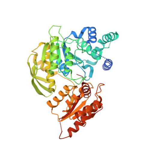
    Folate derivatives are essential vitamins for cell growth and replication, primarily because of their central role in reactions of one-carbon metabolism. Folates require polyglutamation to be efficiently retained within the cell and folate-dependent enzymes have a higher affinity for the polyglutamylated forms of this cofactor. Polyglutamylation is dependent on the enzyme folylpolyglutamate synthetase (FPGS), which catalyzes the sequential addition of several glutamates to folate. FPGS is essential for the growth and survival of important bacterial species, including Mycobacterium tuberculosis, and is a potential drug target. Here, the crystal structures of M. tuberculosis FPGS in complex with ADP and AMPPCP are reported at 2.0 and 2.3 angstroms resolution, respectively. The structures reveal a deeply buried nucleotide-binding site, as in the Escherichia coli and Lactobacillus casei FPGS structures, and a long extended groove for the binding of folate substrates. Differences from the E. coli and L. casei FPGS structures are seen in the binding of a key divalent cation, the carbamylation state of an essential lysine side chain and the adoption of an 'open' position by the active-site beta5-alpha6 loop. These changes point to coordinated events that are associated with dihydropteroate/folate binding and the catalysis of the new amide bond with an incoming glutamate residue.


  • Organizational Affiliation
    • School of Biological Sciences, University of Auckland, Auckland, New Zealand. p.young@auckland.ac.nz

Macromolecule Content 

  • Total Structure Weight: 51.75 kDa 
  • Atom Count: 3,384 
  • Modeled Residue Count: 442 
  • Deposited Residue Count: 487 
  • Unique protein chains: 1

Macromolecules

Find similar proteins by:|  3D Structure
Entity ID: 1
MoleculeChains  Sequence LengthOrganismDetailsImage
FOLYLPOLYGLUTAMATE SYNTHASE PROTEIN FOLC487Mycobacterium tuberculosis H37RvMutation(s): 0 
EC: 6.3.2.17 (PDB Primary Data), 6.3.2.12 (UniProt)
UniProt
Find proteins for O53174 (Mycobacterium tuberculosis (strain CDC 1551 / Oshkosh))
Explore O53174 
Go to UniProtKB:  O53174
Entity Groups
Sequence Clusters30% Identity50% Identity70% Identity90% Identity95% Identity100% Identity
UniProt GroupO53174
Sequence Annotations
Expand
Reference Sequence

Small Molecules

Ligands 3 Unique
IDChains Name / Formula / InChI Key2D Diagram3D Interactions
ACP

Query on ACP


Download:Ideal Coordinates CCD File
B [auth A]PHOSPHOMETHYLPHOSPHONIC ACID ADENYLATE ESTER
C11 H18 N5 O12 P3
UFZTZBNSLXELAL-IOSLPCCCSA-N
GOL

Query on GOL


Download:Ideal Coordinates CCD File
G [auth A],
H [auth A]
GLYCEROL
C3 H8 O3
PEDCQBHIVMGVHV-UHFFFAOYSA-N
CO

Query on CO


Download:Ideal Coordinates CCD File
C [auth A],
D [auth A],
E [auth A],
F [auth A]
COBALT (II) ION
Co
XLJKHNWPARRRJB-UHFFFAOYSA-N

Experimental Data & Validation

Experimental Data

  • Method: X-RAY DIFFRACTION
  • Resolution: 2.30 Å
  • R-Value Free:  0.223 (Depositor), 0.223 (DCC) 
  • R-Value Work:  0.176 (Depositor), 0.175 (DCC) 
  • R-Value Observed: 0.178 (Depositor) 
Space Group: P 21 3
Unit Cell:
Length ( Å )Angle ( ˚ )
a = 112.396α = 90
b = 112.396β = 90
c = 112.396γ = 90
Software Package:
Software NamePurpose
REFMACrefinement
MOSFLMdata reduction
SCALAdata scaling
PHASERphasing

Structure Validation

View Full Validation Report


Ligand Structure Quality Assessment 


Entry History 

Deposition Data

Revision History  (Full details and data files)

  • Version 1.0: 2008-07-01
    Type: Initial release
  • Version 1.1: 2011-07-13
    Changes: Advisory, Refinement description, Version format compliance
  • Version 1.2: 2024-05-01
    Changes: Data collection, Database references, Derived calculations, Other, Refinement description