2W0J | pdb_00002w0j

Crystal structure of Chk2 in complex with NSC 109555, a specific inhibitor


Experimental Data Snapshot

  • Method: X-RAY DIFFRACTION
  • Resolution: 2.05 Å
  • R-Value Free: 
    0.246 (Depositor), 0.255 (DCC) 
  • R-Value Work: 
    0.215 (Depositor), 0.218 (DCC) 
  • R-Value Observed: 
    0.217 (Depositor) 

Starting Model: experimental
View more details

wwPDB Validation 3D Report Full Report

Validation slider image for 2W0J

Ligand Structure Quality Assessment 


This is version 1.3 of the entry. See complete history

Literature

Crystal Structure of Checkpoint Kinase 2 in Complex with Nsc 109555, a Potent and Selective Inhibitor

Lountos, G.T.Tropea, J.E.Zhang, D.Jobson, A.G.Pommier, Y.Shoemaker, R.H.Waugh, D.S.

(2009) Protein Sci 18: 92

  • DOI: https://doi.org/10.1002/pro.16
  • Primary Citation Related Structures: 
    2W0J

  • PubMed Abstract: 

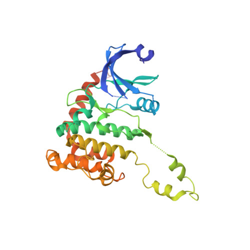
    Checkpoint kinase 2 (Chk2), a ser/thr kinase involved in the ATM-Chk2 checkpoint pathway, is activated by genomic instability and DNA damage and results in either arrest of the cell cycle to allow DNA repair to occur or apoptosis if the DNA damage is severe. Drugs that specifically target Chk2 could be beneficial when administered in combination with current DNA-damaging agents used in cancer therapy. Recently, a novel inhibitor of Chk2, NSC 109555, was identified that exhibited high potency (IC(50) = 240 nM) and selectivity. This compound represents a new chemotype and lead for the development of novel Chk2 inhibitors that could be used as therapeutic agents for the treatment of cancer. To facilitate the discovery of new analogs of NSC 109555 with even greater potency and selectivity, we have solved the crystal structure of this inhibitor in complex with the catalytic domain of Chk2. The structure confirms that the compound is an ATP-competitive inhibitor, as the electron density clearly reveals that it occupies the ATP-binding pocket. However, the mode of inhibition differs from that of the previously studied structure of Chk2 in complex with debromohymenialdisine, a compound that inhibits both Chk1 and Chk2. A unique hydrophobic pocket in Chk2, located very close to the bound inhibitor, presents an opportunity for the rational design of compounds with higher binding affinity and greater selectivity.


  • Organizational Affiliation
    • Macromolecular Crystallography Laboratory, National Cancer Institute at Frederick, P. O. Box B, Frederick, Maryland 21702-1201, USA.

Macromolecule Content 

  • Total Structure Weight: 37.03 kDa 
  • Atom Count: 2,419 
  • Modeled Residue Count: 280 
  • Deposited Residue Count: 323 
  • Unique protein chains: 1

Macromolecules

Find similar proteins by:|  3D Structure
Entity ID: 1
MoleculeChains  Sequence LengthOrganismDetailsImage
SERINE/THREONINE-PROTEIN KINASE CHK2323Homo sapiensMutation(s): 0 
EC: 2.7.11.1
UniProt & NIH Common Fund Data Resources
Find proteins for O96017 (Homo sapiens)
Explore O96017 
Go to UniProtKB:  O96017
PHAROS:  O96017
GTEx:  ENSG00000183765 
Entity Groups
Sequence Clusters30% Identity50% Identity70% Identity90% Identity95% Identity100% Identity
UniProt GroupO96017
Sequence Annotations
Expand
Reference Sequence

Small Molecules

Ligands 2 Unique
IDChains Name / Formula / InChI Key2D Diagram3D Interactions
ZAT

Query on ZAT


Download:Ideal Coordinates CCD File
B [auth A]4,4'-DIACETYLDIPHENYLUREA-BIS(GUANYLHYDRAZONE)
C19 H24 N10 O
HCAQGQIHBFVVIX-LYXAAFRTSA-N
NO3

Query on NO3


Download:Ideal Coordinates CCD File
C [auth A]NITRATE ION
N O3
NHNBFGGVMKEFGY-UHFFFAOYSA-N

Experimental Data & Validation

Experimental Data

  • Method: X-RAY DIFFRACTION
  • Resolution: 2.05 Å
  • R-Value Free:  0.246 (Depositor), 0.255 (DCC) 
  • R-Value Work:  0.215 (Depositor), 0.218 (DCC) 
  • R-Value Observed: 0.217 (Depositor) 
Space Group: P 32 2 1
Unit Cell:
Length ( Å )Angle ( ˚ )
a = 90.847α = 90
b = 90.847β = 90
c = 93.526γ = 120
Software Package:
Software NamePurpose
REFMACrefinement
HKL-3000data reduction
SCALEPACKdata scaling
MOLREPphasing

Structure Validation

View Full Validation Report


Ligand Structure Quality Assessment 


Entry History 

Deposition Data

Revision History  (Full details and data files)

  • Version 1.0: 2009-02-10
    Type: Initial release
  • Version 1.1: 2011-05-08
    Changes: Version format compliance
  • Version 1.2: 2011-07-13
    Changes: Version format compliance
  • Version 1.3: 2023-12-13
    Changes: Data collection, Database references, Other, Refinement description