2W8Y

RU486 bound to the progesterone receptor in a destabilized agonistic conformation


Experimental Data Snapshot

  • Method: X-RAY DIFFRACTION
  • Resolution: 1.80 Å
  • R-Value Free: 0.218 
  • R-Value Work: 0.185 
  • R-Value Observed: 0.187 

wwPDB Validation   3D Report Full Report


Ligand Structure Quality Assessment 


This is version 1.2 of the entry. See complete history


Literature

The X-Ray Structure of Ru486 Bound to the Progesterone Receptor in a Destabilized Agonistic Conformation.

Raaijmakers, H.C.A.Versteegh, J.Uitdehaag, J.C.M.

(2009) J Biol Chem 284: 19572

  • DOI: https://doi.org/10.1074/jbc.M109.007872
  • Primary Citation of Related Structures:  
    2W8Y

  • PubMed Abstract: 

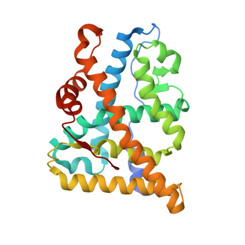
    Here we describe the 1.95 A structure of the clinically used antiprogestin RU486 (mifepristone) in complex with the progesterone receptor (PR). The structure was obtained by taking a crystal of the PR ligand binding domain containing the agonist norethindrone and soaking it in a solution containing the antagonist RU486 for extended times. Clear ligand exchange could be observed in one copy of the PR ligand binding domain dimer in the crystal. RU486 binds while PR is in an agonistic conformation without displacing helix 12. Although this is probably because of the constraints of the crystal lattice, it demonstrates that helix 12 displacement is not a prerequisite for RU486 binding. Interestingly, B-factor analysis clearly shows that helix 12 becomes more flexible after RU486 binding, suggesting that RU486, being a model antagonist, does not induce one fixed conformation of helix 12 but changes its positional equilibrium. This conclusion is confirmed by comparing the structures of RU486 bound to PR and RU486 bound to the glucocorticoid receptor.


  • Organizational Affiliation

    Departments of Molecular Design and Informatics, Molecular Pharmacology and DMPK, Schering-Plough Research Institute, P. O. Box 20, 5340 BH Oss, The Netherlands.


Macromolecules
Find similar proteins by:  (by identity cutoff)  |  3D Structure
Entity ID: 1
MoleculeChains Sequence LengthOrganismDetailsImage
PROGESTERONE RECEPTOR
A, B
260Homo sapiensMutation(s): 0 
UniProt & NIH Common Fund Data Resources
Find proteins for P06401 (Homo sapiens)
Explore P06401 
Go to UniProtKB:  P06401
PHAROS:  P06401
GTEx:  ENSG00000082175 
Entity Groups  
Sequence Clusters30% Identity50% Identity70% Identity90% Identity95% Identity100% Identity
UniProt GroupP06401
Sequence Annotations
Expand
  • Reference Sequence
Small Molecules
Ligands 4 Unique
IDChains Name / Formula / InChI Key2D Diagram3D Interactions
486
Query on 486

Download Ideal Coordinates CCD File 
C [auth A]11-(4-DIMETHYLAMINO-PHENYL)-17-HYDROXY-13-METHYL-17-PROP-1-YNYL-1,2,6,7,8,11,12,13,14,15,16,17-DODEC AHYDRO-CYCLOPENTA[A]PHENANTHREN-3-ONE
C29 H35 N O2
VKHAHZOOUSRJNA-GCNJZUOMSA-N
NDR
Query on NDR

Download Ideal Coordinates CCD File 
E [auth B](14beta,17alpha)-17-ethynyl-17-hydroxyestr-4-en-3-one
C20 H26 O2
VIKNJXKGJWUCNN-XGXHKTLJSA-N
SO4
Query on SO4

Download Ideal Coordinates CCD File 
D [auth A]SULFATE ION
O4 S
QAOWNCQODCNURD-UHFFFAOYSA-L
EDO
Query on EDO

Download Ideal Coordinates CCD File 
F [auth B]1,2-ETHANEDIOL
C2 H6 O2
LYCAIKOWRPUZTN-UHFFFAOYSA-N
Binding Affinity Annotations 
IDSourceBinding Affinity
486 BindingDB:  2W8Y Ki: min: 0.25, max: 15 (nM) from 7 assay(s)
IC50: min: 0.02, max: 10 (nM) from 25 assay(s)
EC50: min: 0.3, max: 16 (nM) from 2 assay(s)
PDBBind:  2W8Y Ki: 0.82 (nM) from 1 assay(s)
NDR BindingDB:  2W8Y Ki: 1.9 (nM) from 1 assay(s)
Kd: 0.4 (nM) from 1 assay(s)
IC50: 3.2 (nM) from 1 assay(s)
EC50: 2.2 (nM) from 1 assay(s)
Experimental Data & Validation

Experimental Data

  • Method: X-RAY DIFFRACTION
  • Resolution: 1.80 Å
  • R-Value Free: 0.218 
  • R-Value Work: 0.185 
  • R-Value Observed: 0.187 
  • Space Group: P 1 21 1
Unit Cell:
Length ( Å )Angle ( ˚ )
a = 58.164α = 90
b = 63.897β = 95.57
c = 70.051γ = 90
Software Package:
Software NamePurpose
REFMACrefinement
MOSFLMdata reduction
SCALAdata scaling

Structure Validation

View Full Validation Report


Ligand Structure Quality Assessment 


Entry History 

Deposition Data

Revision History  (Full details and data files)

  • Version 1.0: 2009-04-28
    Type: Initial release
  • Version 1.1: 2011-07-13
    Changes: Advisory, Refinement description, Version format compliance
  • Version 1.2: 2017-12-27
    Changes: Advisory, Structure summary