2WKO

Structure of metal loaded Pathogenic SOD1 Mutant G93A.


Experimental Data Snapshot

  • Method: X-RAY DIFFRACTION
  • Resolution: 1.97 Å
  • R-Value Free: 0.231 
  • R-Value Work: 0.174 
  • R-Value Observed: 0.177 

wwPDB Validation   3D Report Full Report


This is version 1.3 of the entry. See complete history


Literature

Structural and Biophysical Properties of Metal-Free Pathogenic Sod1 Mutants A4V and G93A.

Galaleldeen, A.Strange, R.W.Whitson, L.J.Antonyuk, S.V.Narayana, N.Taylor, A.B.Schuermann, J.P.Holloway, S.P.Hasnain, S.S.Hart, P.J.

(2009) Arch Biochem Biophys 492: 40

  • DOI: https://doi.org/10.1016/j.abb.2009.09.020
  • Primary Citation of Related Structures:  
    2WKO, 3GZO, 3GZP, 3GZQ

  • PubMed Abstract: 

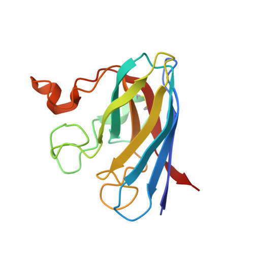
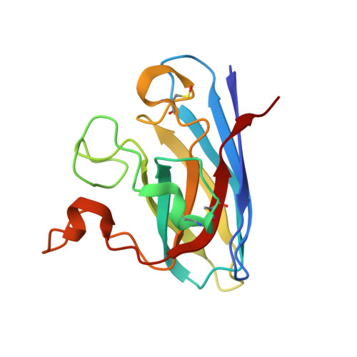
    Amyotrophic lateral sclerosis (ALS) is a fatal, progressive neurodegenerative disease characterized by the destruction of motor neurons in the spinal cord and brain. A subset of ALS cases are linked to dominant mutations in copper-zinc superoxide dismutase (SOD1). The pathogenic SOD1 variants A4V and G93A have been the foci of multiple studies aimed at understanding the molecular basis for SOD1-linked ALS. The A4V variant is responsible for the majority of familial ALS cases in North America, causing rapidly progressing paralysis once symptoms begin and the G93A SOD1 variant is overexpressed in often studied murine models of the disease. Here we report the three-dimensional structures of metal-free A4V and of metal-bound and metal-free G93A SOD1. In the metal-free structures, the metal-binding loop elements are observed to be severely disordered, suggesting that these variants may share mechanisms of aggregation proposed previously for other pathogenic SOD1 proteins.


  • Organizational Affiliation

    Department of Biochemistry and the X-ray Crystallography Core Laboratory, The University of Texas Health Science Center at San Antonio, 7703 Floyd Curl Drive, San Antonio, TX 78229-3900, USA.


Macromolecules
Find similar proteins by:  (by identity cutoff)  |  3D Structure
Entity ID: 1
MoleculeChains Sequence LengthOrganismDetailsImage
SUPEROXIDE DISMUTASE [CU-ZN]154Homo sapiensMutation(s): 1 
EC: 1.15.1.1
UniProt & NIH Common Fund Data Resources
Find proteins for P00441 (Homo sapiens)
Explore P00441 
Go to UniProtKB:  P00441
PHAROS:  P00441
GTEx:  ENSG00000142168 
Entity Groups  
Sequence Clusters30% Identity50% Identity70% Identity90% Identity95% Identity100% Identity
UniProt GroupP00441
Sequence Annotations
Expand
  • Reference Sequence
Find similar proteins by:  (by identity cutoff)  |  3D Structure
Entity ID: 2
MoleculeChains Sequence LengthOrganismDetailsImage
SUPEROXIDE DISMUTASE [CU-ZN]B [auth F]154Homo sapiensMutation(s): 1 
EC: 1.15.1.1
UniProt & NIH Common Fund Data Resources
Find proteins for P00441 (Homo sapiens)
Explore P00441 
Go to UniProtKB:  P00441
PHAROS:  P00441
GTEx:  ENSG00000142168 
Entity Groups  
Sequence Clusters30% Identity50% Identity70% Identity90% Identity95% Identity100% Identity
UniProt GroupP00441
Sequence Annotations
Expand
  • Reference Sequence
Experimental Data & Validation

Experimental Data

  • Method: X-RAY DIFFRACTION
  • Resolution: 1.97 Å
  • R-Value Free: 0.231 
  • R-Value Work: 0.174 
  • R-Value Observed: 0.177 
  • Space Group: P 1 21 1
Unit Cell:
Length ( Å )Angle ( ˚ )
a = 51.488α = 90
b = 47.157β = 90.38
c = 56.356γ = 90
Software Package:
Software NamePurpose
REFMACrefinement
HKL-2000data reduction
HKL-2000data scaling
MOLREPphasing

Structure Validation

View Full Validation Report


Entry History 

Deposition Data

Revision History  (Full details and data files)

  • Version 1.0: 2009-11-24
    Type: Initial release
  • Version 1.1: 2011-05-08
    Changes: Version format compliance
  • Version 1.2: 2011-07-13
    Changes: Version format compliance
  • Version 1.3: 2023-12-13
    Changes: Data collection, Database references, Derived calculations, Other, Refinement description