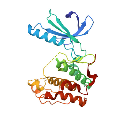
2WQO

STRUCTURE OF NEK2 BOUND TO THE AMINOPYRIDINE CCT241950


Experimental Data Snapshot

  • Method: X-RAY DIFFRACTION
  • Resolution: 2.17 Å
  • R-Value Free: 0.235 
  • R-Value Work: 0.183 
  • R-Value Observed: 0.186 

wwPDB Validation   3D Report Full Report


Ligand Structure Quality Assessment 


This is version 1.3 of the entry. See complete history


Literature

An Autoinhibitory Tyrosine Motif in the Cell-Cycle- Regulated Nek7 Kinase is Released Through Binding of Nek9.

Richards, M.W.O'Regan, L.Mas-Droux, C.Blot, J.M.Cheung, J.Hoelder, S.Fry, A.M.Bayliss, R.

(2009) Mol Cell 36: 560

  • DOI: https://doi.org/10.1016/j.molcel.2009.09.038
  • Primary Citation of Related Structures:  
    2WQM, 2WQN, 2WQO

  • PubMed Abstract: 

    Mitosis is controlled by multiple protein kinases, many of which are abnormally expressed in human cancers. Nek2, Nek6, Nek7, and Nek9 are NIMA-related kinases essential for proper mitotic progression. We determined the atomic structure of Nek7 and discovered an autoinhibited conformation that suggests a regulatory mechanism not previously described in kinases. Additionally, Nek2 adopts the same conformation when bound to a drug-like molecule. In both structures, a tyrosine side chain points into the active site, interacts with the activation loop, and blocks the alphaC helix. Tyrosine mutants of Nek7 and the related kinase Nek6 are constitutively active. The activity of Nek6 and Nek7, but not the tyrosine mutant, is increased by interaction with the Nek9 noncatalytic C-terminal domain, suggesting a mechanism in which the tyrosine is released from its autoinhibitory position. The autoinhibitory conformation is common to three Neks and provides a potential target for selective kinase inhibitors.


  • Organizational Affiliation

    Institute of Cancer Research, London, UK.


Macromolecules
Find similar proteins by:  (by identity cutoff)  |  3D Structure
Entity ID: 1
MoleculeChains Sequence LengthOrganismDetailsImage
SERINE/THREONINE-PROTEIN KINASE NEK2279Homo sapiensMutation(s): 0 
EC: 2.7.11.1
UniProt & NIH Common Fund Data Resources
Find proteins for P51955 (Homo sapiens)
Explore P51955 
Go to UniProtKB:  P51955
PHAROS:  P51955
GTEx:  ENSG00000117650 
Entity Groups  
Sequence Clusters30% Identity50% Identity70% Identity90% Identity95% Identity100% Identity
UniProt GroupP51955
Sequence Annotations
Expand
  • Reference Sequence
Experimental Data & Validation

Experimental Data

  • Method: X-RAY DIFFRACTION
  • Resolution: 2.17 Å
  • R-Value Free: 0.235 
  • R-Value Work: 0.183 
  • R-Value Observed: 0.186 
  • Space Group: C 1 2 1
Unit Cell:
Length ( Å )Angle ( ˚ )
a = 100.43α = 90
b = 56.84β = 128.09
c = 73.49γ = 90
Software Package:
Software NamePurpose
PHENIXrefinement
MOSFLMdata reduction
SCALAdata scaling
PHENIXphasing

Structure Validation

View Full Validation Report


Ligand Structure Quality Assessment 


Entry History 

Deposition Data

Revision History  (Full details and data files)

  • Version 1.0: 2009-12-08
    Type: Initial release
  • Version 1.1: 2011-05-08
    Changes: Version format compliance
  • Version 1.2: 2011-07-13
    Changes: Version format compliance
  • Version 1.3: 2024-05-08
    Changes: Data collection, Database references, Derived calculations, Other