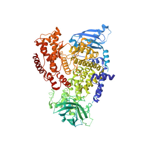
2WXK

The crystal structure of the murine class IA PI 3-kinase p110delta in complex with INK666.


Experimental Data Snapshot

  • Method: X-RAY DIFFRACTION
  • Resolution: 2.90 Å
  • R-Value Free: 0.287 
  • R-Value Work: 0.213 
  • R-Value Observed: 0.215 

Starting Model: experimental
View more details

wwPDB Validation   3D Report Full Report


Ligand Structure Quality Assessment 


This is version 1.3 of the entry. See complete history


Literature

The P110D Structure: Mechanisms for Selectivity and Potency of New Pi(3)K Inhibitors

Berndt, A.Miller, S.Williams, O.Lee, D.D.Houseman, B.T.Pacold, J.I.Gorrec, F.Hon, W.-C.Liu, Y.Rommel, C.Gaillard, P.Ruckle, T.Schwarz, M.K.Shokat, K.M.Shaw, J.P.Williams, R.L.

(2010) Nat Chem Biol 6: 117

  • DOI: https://doi.org/10.1038/nchembio.293
  • Primary Citation of Related Structures:  
    2WXF, 2WXG, 2WXH, 2WXI, 2WXJ, 2WXK, 2WXL, 2WXM, 2WXN, 2WXO, 2WXP, 2WXQ, 2WXR, 2X38

  • PubMed Abstract: 

    Deregulation of the phosphoinositide-3-OH kinase (PI(3)K) pathway has been implicated in numerous pathologies including cancer, diabetes, thrombosis, rheumatoid arthritis and asthma. Recently, small-molecule and ATP-competitive PI(3)K inhibitors with a wide range of selectivities have entered clinical development. In order to understand the mechanisms underlying the isoform selectivity of these inhibitors, we developed a new expression strategy that enabled us to determine to our knowledge the first crystal structure of the catalytic subunit of the class IA PI(3)K p110 delta. Structures of this enzyme in complex with a broad panel of isoform- and pan-selective class I PI(3)K inhibitors reveal that selectivity toward p110 delta can be achieved by exploiting its conformational flexibility and the sequence diversity of active site residues that do not contact ATP. We have used these observations to rationalize and synthesize highly selective inhibitors for p110 delta with greatly improved potencies.


  • Organizational Affiliation

    Medical Research Council-Laboratory of Molecular Biology, Cambridge, UK.


Macromolecules
Find similar proteins by:  (by identity cutoff)  |  3D Structure
Entity ID: 1
MoleculeChains Sequence LengthOrganismDetailsImage
PHOSPHATIDYLINOSITOL-4,5-BISPHOSPHATE 3-KINASE CATALYTIC SUBUNIT DELTA ISOFORM940Mus musculusMutation(s): 0 
EC: 2.7.1.153 (PDB Primary Data), 2.7.1.137 (UniProt)
UniProt
Find proteins for O35904 (Mus musculus)
Explore O35904 
Go to UniProtKB:  O35904
Entity Groups  
Sequence Clusters30% Identity50% Identity70% Identity90% Identity95% Identity100% Identity
UniProt GroupO35904
Sequence Annotations
Expand
  • Reference Sequence
Small Molecules
Ligands 1 Unique
IDChains Name / Formula / InChI Key2D Diagram3D Interactions
RW4
Query on RW4

Download Ideal Coordinates CCD File 
B [auth A]3-(2-amino-1,3-benzothiazol-6-yl)-1-{[2-(4-methylpiperazin-1-yl)quinolin-3-yl]methyl}-1H-pyrazolo[3,4-d]pyrimidin-4-amine
C27 H26 N10 S
UNSGOTITAWKECZ-UHFFFAOYSA-N
Binding Affinity Annotations 
IDSourceBinding Affinity
RW4 BindingDB:  2WXK IC50: 0.5 (nM) from 1 assay(s)
PDBBind:  2WXK IC50: 0.5 (nM) from 1 assay(s)
Experimental Data & Validation

Experimental Data

  • Method: X-RAY DIFFRACTION
  • Resolution: 2.90 Å
  • R-Value Free: 0.287 
  • R-Value Work: 0.213 
  • R-Value Observed: 0.215 
  • Space Group: C 2 2 21
Unit Cell:
Length ( Å )Angle ( ˚ )
a = 64.362α = 90
b = 143.669β = 90
c = 223.121γ = 90
Software Package:
Software NamePurpose
REFMACrefinement
iMOSFLMdata reduction
SCALAdata scaling
PHASERphasing

Structure Validation

View Full Validation Report


Ligand Structure Quality Assessment 


Entry History 

Revision History  (Full details and data files)

  • Version 1.0: 2010-01-12
    Type: Initial release
  • Version 1.1: 2011-07-13
    Changes: Advisory, Refinement description, Version format compliance
  • Version 1.2: 2019-04-24
    Changes: Data collection, Other, Source and taxonomy
  • Version 1.3: 2023-12-20
    Changes: Data collection, Database references, Derived calculations, Other, Refinement description