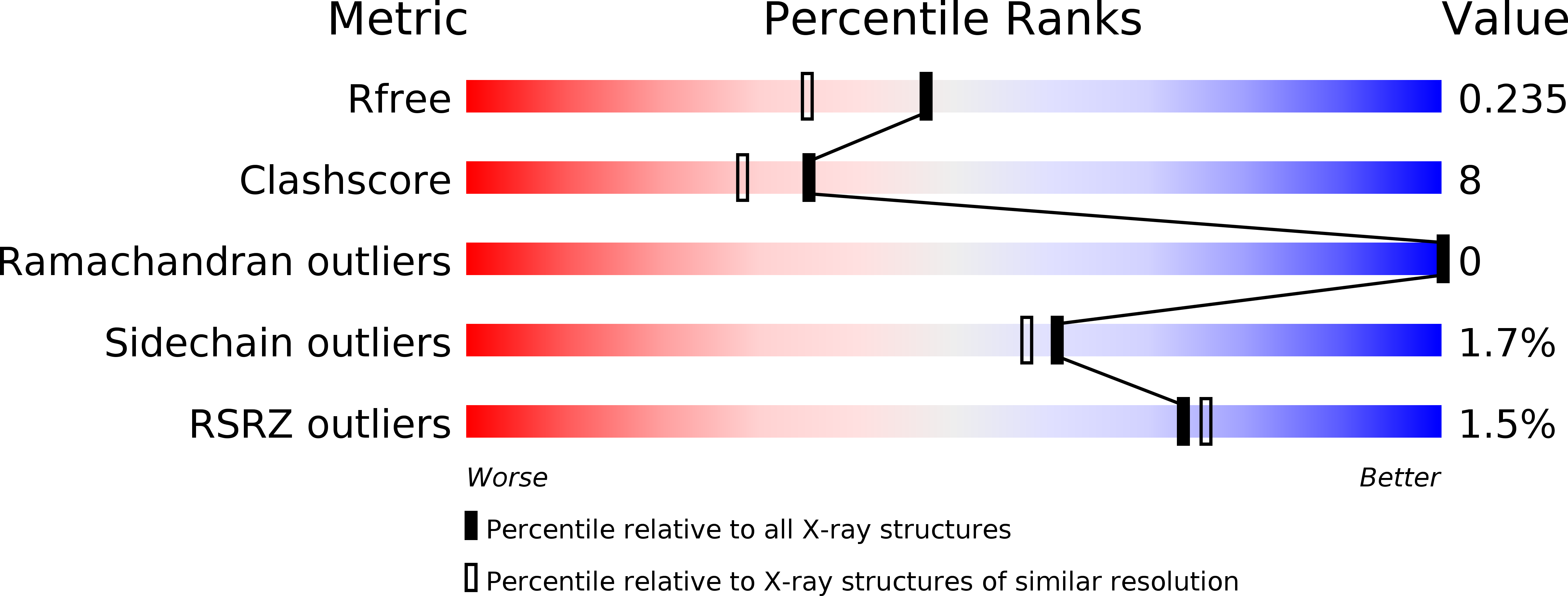
2X34

Structure of a polyisoprenoid binding domain from Saccharophagus degradans implicated in plant cell wall breakdown


Experimental Data Snapshot

  • Method: X-RAY DIFFRACTION
  • Resolution: 1.90 Å
  • R-Value Free: 
    0.223 (Depositor), 0.235 (DCC) 
  • R-Value Work: 
    0.175 (Depositor), 0.190 (DCC) 
  • R-Value Observed: 
    0.178 (Depositor) 

wwPDB Validation   3D Report Full Report


Ligand Structure Quality Assessment 

Created with Raphaël 2.3.0Worse 01 BetterLigand structure goodness of fit to experimental dataBest fitted UQ8Click on this verticalbar to view details

This is version 1.3 of the entry. See complete history


Literature

Structure of a Polyisoprenoid Binding Domain from Saccharophagus Degradans Implicated in Plant Cell Wall Breakdown

Vincent, F.Dalmolin, D.Weiner, R.M.Bourne, Y.Henrissat, B.

(2010) FEBS Lett 584: 1577

  • DOI: https://doi.org/10.1016/j.febslet.2010.03.015
  • Primary Citation of Related Structures:  
    2X32, 2X34

  • PubMed Abstract: 

    Saccharophagus degradans belongs to a recently discovered group of marine bacteria equipped with an arsenal of sugar cleaving enzymes coupled to carbohydrate-binding domains to degrade various insoluble complex polysaccharides. The modular Sde-1182 protein consists of a family 2 carbohydrate binding module linked to a X158 domain of unknown function. The 1.9 A and 1.55 A resolution crystal structures of the isolated X158 domain bound to the two related polyisoprenoid molecules, ubiquinone and octaprenyl pyrophosphate, unveil a beta-barrel architecture reminiscent of the YceI-like superfamily that resembles the architecture of the lipocalin fold. This unprecedented association coupling oxidoreduction and carbohydrate recognition events may have implications for effective nutrient uptake in the marine environment.


  • Organizational Affiliation

    Architecture et Fonction des Macromolécules Biologiques, UMR6098, CNRS and Aix-Marseille Universities, Marseille, France. florence.vincent@afmb.univ-mrs.fr


Macromolecules
Find similar proteins by:  (by identity cutoff)  |  3D Structure
Entity ID: 1
MoleculeChains Sequence LengthOrganismDetailsImage
CELLULOSE-BINDING PROTEIN, X158
A, B
181Saccharophagus degradans 2-40Mutation(s): 0 
UniProt
Find proteins for Q21LI5 (Saccharophagus degradans (strain 2-40 / ATCC 43961 / DSM 17024))
Explore Q21LI5 
Go to UniProtKB:  Q21LI5
Entity Groups  
Sequence Clusters30% Identity50% Identity70% Identity90% Identity95% Identity100% Identity
UniProt GroupQ21LI5
Sequence Annotations
Expand
  • Reference Sequence
Small Molecules
Modified Residues  1 Unique
IDChains TypeFormula2D DiagramParent
MSE
Query on MSE
A, B
L-PEPTIDE LINKINGC5 H11 N O2 SeMET
Experimental Data & Validation

Experimental Data

  • Method: X-RAY DIFFRACTION
  • Resolution: 1.90 Å
  • R-Value Free:  0.223 (Depositor), 0.235 (DCC) 
  • R-Value Work:  0.175 (Depositor), 0.190 (DCC) 
  • R-Value Observed: 0.178 (Depositor) 
Space Group: P 21 21 21
Unit Cell:
Length ( Å )Angle ( ˚ )
a = 62.979α = 90
b = 64.964β = 90
c = 84.191γ = 90
Software Package:
Software NamePurpose
REFMACrefinement
MOSFLMdata reduction
SCALAdata scaling
AutoSolphasing

Structure Validation

View Full Validation Report


Ligand Structure Quality Assessment 

Created with Raphaël 2.3.0Worse 01 BetterLigand structure goodness of fit to experimental dataBest fitted UQ8Click on this verticalbar to view details

Entry History 

Deposition Data

Revision History  (Full details and data files)

  • Version 1.0: 2010-03-23
    Type: Initial release
  • Version 1.1: 2011-07-13
    Changes: Advisory, Version format compliance
  • Version 1.2: 2018-03-07
    Changes: Advisory, Source and taxonomy
  • Version 1.3: 2024-10-23
    Changes: Advisory, Data collection, Database references, Derived calculations, Other, Structure summary