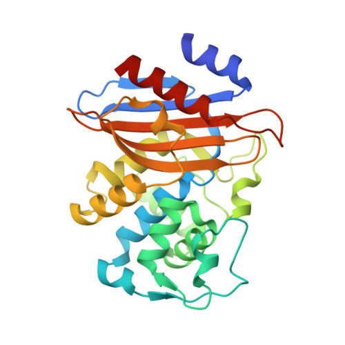
2X71

Structural basis for the interaction of lactivicins with serine beta- lactamases


Experimental Data Snapshot

  • Method: X-RAY DIFFRACTION
  • Resolution: 2.10 Å
  • R-Value Free: 0.227 
  • R-Value Work: 0.187 
  • R-Value Observed: 0.189 

Starting Model: experimental
View more details

wwPDB Validation   3D Report Full Report


Ligand Structure Quality Assessment 


This is version 1.3 of the entry. See complete history


Literature

Structural Basis for the Interaction of Lactivicins with Serine Beta-Lactamases.

Brown, T.Charlier, P.Herman, R.Schofield, C.J.Sauvage, E.

(2010) J Med Chem 53: 5890

  • DOI: https://doi.org/10.1021/jm100437u
  • Primary Citation of Related Structures:  
    2X71

  • PubMed Abstract: 

    Lactivicin (LTV) is a natural non-beta-lactam antibiotic that inhibits penicillin-binding proteins and serine beta-lactamases. A crystal structure of a BS3-LTV complex reveals that, as for its reaction with PBPs, LTV reacts with the nucleophilic serine and that cycloserine and lactone rings of LTV are opened. This structure, together with reported structures of PBP1b with lactivicins, provides a basis for developing improved lactivicin-based gamma-lactam antibiotics.


  • Organizational Affiliation

    Chemistry Research Laboratory, Department of Chemistry, University of Oxford, Oxford, UK.


Macromolecules
Find similar proteins by:  (by identity cutoff)  |  3D Structure
Entity ID: 1
MoleculeChains Sequence LengthOrganismDetailsImage
BETA-LACTAMASE
A, B
265Bacillus licheniformisMutation(s): 0 
EC: 3.5.2.6
UniProt
Find proteins for P94458 (Bacillus licheniformis)
Explore P94458 
Go to UniProtKB:  P94458
Entity Groups  
Sequence Clusters30% Identity50% Identity70% Identity90% Identity95% Identity100% Identity
UniProt GroupP94458
Sequence Annotations
Expand
  • Reference Sequence
Experimental Data & Validation

Experimental Data

  • Method: X-RAY DIFFRACTION
  • Resolution: 2.10 Å
  • R-Value Free: 0.227 
  • R-Value Work: 0.187 
  • R-Value Observed: 0.189 
  • Space Group: P 1 21 1
Unit Cell:
Length ( Å )Angle ( ˚ )
a = 46.652α = 90
b = 104.089β = 94.45
c = 62.838γ = 90
Software Package:
Software NamePurpose
REFMACrefinement
MOSFLMdata reduction
SCALAdata scaling

Structure Validation

View Full Validation Report


Ligand Structure Quality Assessment 


Entry History 

Deposition Data

Revision History  (Full details and data files)

  • Version 1.0: 2010-07-14
    Type: Initial release
  • Version 1.1: 2011-07-13
    Changes: Advisory, Version format compliance
  • Version 1.2: 2011-11-02
    Changes: Database references
  • Version 1.3: 2023-12-20
    Changes: Data collection, Database references, Derived calculations, Other, Refinement description