2XR5

Crystal structure of the complex of the carbohydrate recognition domain of human DC-SIGN with pseudo dimannoside mimic.


Experimental Data Snapshot

  • Method: X-RAY DIFFRACTION
  • Resolution: 1.42 Å
  • R-Value Free: 0.171 
  • R-Value Work: 0.148 
  • R-Value Observed: 0.150 

wwPDB Validation   3D Report Full Report


Ligand Structure Quality Assessment 


This is version 2.1 of the entry. See complete history


Literature

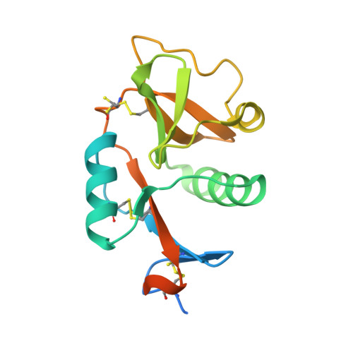
Structure of a Glycomimetic Ligand in the Carbohydrate Recognition Domain of C-Type Lectin Dc-Sign. Structural Requirements for Selectivity and Ligand Design.

Thepaut, M.Guzzi, C.Sutkeviciute, I.Sattin, S.Ribeiro-Viana, R.Varga, N.Chabrol, E.Rojo, J.Bernardi, A.Angulo, J.Nieto, P.M.Fieschi, F.

(2013) J Am Chem Soc 135: 2518

  • DOI: https://doi.org/10.1021/ja3053305
  • Primary Citation of Related Structures:  
    2XR5

  • PubMed Abstract: 

    In genital mucosa, different fates are described for HIV according to the subtype of dendritic cells (DCs) involved in its recognition. This notably depends on the C-type lectin receptor, langerin or DC-SIGN, involved in gp120 interaction. Langerin blocks HIV transmission by its internalization in specific organelles of Langerhans cells. On the contrary, DC-SIGN enhances HIV trans-infection of T lymphocytes. Thus, approaches aiming to inhibit DC-SIGN, without blocking langerin, represent attractive anti-HIV strategies. We previously demonstrated that dendrons bearing multiple copies of glycomimetic compounds were able to block DC-SIGN-dependent HIV infection in cervical explant models. Optimization of such ligand requires detailed characterization of its binding mode. In the present work, we determined the first high-resolution structure of a glycomimetic/DC-SIGN complex by X-ray crystallography. This glycomimetic, pseudo-1,2-mannobioside, shares shape and conformational properties with Manα1-2Man, its natural counterpart. However, it uses the binding epitope previously described for Lewis X, a ligand specific for DC-SIGN among the C-type lectin family. Thus, selectivity gain for DC-SIGN versus langerin is observed with pseudo-1,2-mannobioside as shown by surface plasmon resonance analysis. In parallel, ligand binding was also analyzed by TR-NOESY and STD NMR experiments, combined with the CORCEMA-ST protocol. These studies demonstrate that the complex, defined by X-ray crystallography, represents the unique binding mode of this ligand as opposed to the several binding orientations described for the natural ligand. This exclusive binding mode and its selective interaction properties position this glycomimetic as a good lead compound for rational improvement based on a structurally driven approach.


  • Organizational Affiliation

    Institut de Biologie Structurale, Université Grenoble I, 41 rue Jules Horowitz, Grenoble, F-38027, France.


Macromolecules
Find similar proteins by:  (by identity cutoff)  |  3D Structure
Entity ID: 1
MoleculeChains Sequence LengthOrganismDetailsImage
CD209 ANTIGEN166Homo sapiensMutation(s): 0 
UniProt & NIH Common Fund Data Resources
Find proteins for Q9NNX6 (Homo sapiens)
Explore Q9NNX6 
Go to UniProtKB:  Q9NNX6
PHAROS:  Q9NNX6
GTEx:  ENSG00000090659 
Entity Groups  
Sequence Clusters30% Identity50% Identity70% Identity90% Identity95% Identity100% Identity
UniProt GroupQ9NNX6
Sequence Annotations
Expand
  • Reference Sequence
Small Molecules
Ligands 5 Unique
IDChains Name / Formula / InChI Key2D Diagram3D Interactions
07B
Query on 07B

Download Ideal Coordinates CCD File 
C [auth A]dimethyl (1S,2S,4S,5S)-4,5-dihydroxycyclohexane-1,2-dicarboxylate
C10 H16 O6
ICVNZKRWDVXSLF-XAMCCFCMSA-N
MAN
Query on MAN

Download Ideal Coordinates CCD File 
B [auth A]alpha-D-mannopyranose
C6 H12 O6
WQZGKKKJIJFFOK-PQMKYFCFSA-N
ETA
Query on ETA

Download Ideal Coordinates CCD File 
D [auth A]ETHANOLAMINE
C2 H7 N O
HZAXFHJVJLSVMW-UHFFFAOYSA-N
CA
Query on CA

Download Ideal Coordinates CCD File 
E [auth A],
F [auth A],
G [auth A]
CALCIUM ION
Ca
BHPQYMZQTOCNFJ-UHFFFAOYSA-N
CL
Query on CL

Download Ideal Coordinates CCD File 
H [auth A],
I [auth A],
J [auth A]
CHLORIDE ION
Cl
VEXZGXHMUGYJMC-UHFFFAOYSA-M
Experimental Data & Validation

Experimental Data

  • Method: X-RAY DIFFRACTION
  • Resolution: 1.42 Å
  • R-Value Free: 0.171 
  • R-Value Work: 0.148 
  • R-Value Observed: 0.150 
  • Space Group: P 43 21 2
Unit Cell:
Length ( Å )Angle ( ˚ )
a = 71.45α = 90
b = 71.45β = 90
c = 52.67γ = 90
Software Package:
Software NamePurpose
REFMACrefinement
XDSdata reduction
XSCALEdata scaling
MOLREPphasing

Structure Validation

View Full Validation Report


Ligand Structure Quality Assessment 


Entry History 

Deposition Data

Revision History  (Full details and data files)

  • Version 1.0: 2011-10-19
    Type: Initial release
  • Version 1.1: 2013-12-25
    Changes: Database references
  • Version 1.2: 2019-01-30
    Changes: Data collection, Experimental preparation, Other
  • Version 1.3: 2019-03-06
    Changes: Data collection, Derived calculations, Experimental preparation
  • Version 1.4: 2020-07-29
    Type: Remediation
    Reason: Carbohydrate remediation
    Changes: Data collection, Derived calculations, Other, Structure summary
  • Version 2.0: 2023-11-15
    Changes: Atomic model, Data collection, Database references, Derived calculations, Structure summary
  • Version 2.1: 2023-12-20
    Changes: Refinement description