2XTT

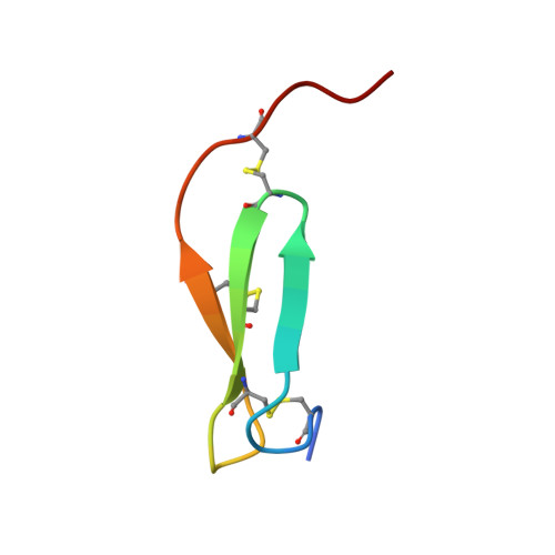
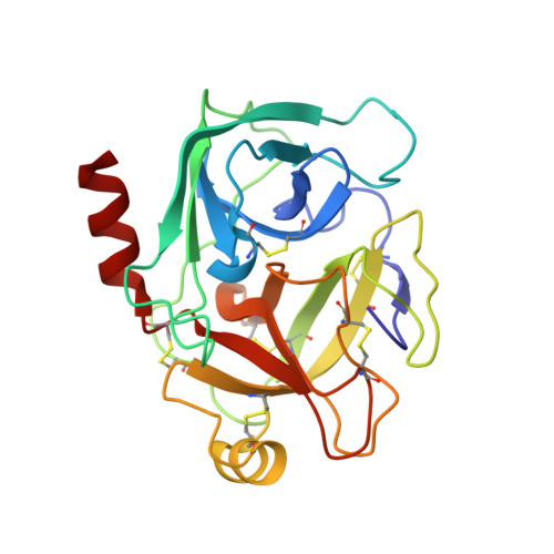
Bovine trypsin in complex with evolutionary enhanced Schistocerca gregaria protease inhibitor 1 (SGPI-1-P02)


Experimental Data Snapshot

  • Method: X-RAY DIFFRACTION
  • Resolution: 0.93 Å
  • R-Value Free: 0.140 

Starting Model: experimental
View more details

wwPDB Validation   3D Report Full Report


This is version 1.7 of the entry. See complete history


Literature

The catalytic aspartate is protonated in the Michaelis complex formed between trypsin and an in vitro evolved substrate-like inhibitor: a refined mechanism of serine protease action.

Wahlgren, W.Y.Pal, G.Kardos, J.Porrogi, P.Szenthe, B.Patthy, A.Graf, L.Katona, G.

(2011) J Biol Chem 286: 3587-3596

  • DOI: https://doi.org/10.1074/jbc.M110.161604
  • Primary Citation of Related Structures:  
    2XTT

  • PubMed Abstract: 

    The mechanism of serine proteases prominently illustrates how charged amino acid residues and proton transfer events facilitate enzyme catalysis. Here we present an ultrahigh resolution (0.93 Å) x-ray structure of a complex formed between trypsin and a canonical inhibitor acting through a substrate-like mechanism. The electron density indicates the protonation state of all catalytic residues where the catalytic histidine is, as expected, in its neutral state prior to the acylation step by the catalytic serine. The carboxyl group of the catalytic aspartate displays an asymmetric electron density so that the O(δ2)-C(γ) bond appears to be a double bond, with O(δ2) involved in a hydrogen bond to His-57 and Ser-214. Only when Asp-102 is protonated on O(δ1) atom could a density functional theory simulation reproduce the observed electron density. The presence of a putative hydrogen atom is also confirmed by a residual mF(obs) - DF(calc) density above 2.5 σ next to O(δ1). As a possible functional role for the neutral aspartate in the active site, we propose that in the substrate-bound form, the neutral aspartate residue helps to keep the pK(a) of the histidine sufficiently low, in the active neutral form. When the histidine receives a proton during the catalytic cycle, the aspartate becomes simultaneously negatively charged, providing additional stabilization for the protonated histidine and indirectly to the tetrahedral intermediate. This novel proposal unifies the seemingly conflicting experimental observations, which were previously seen as either supporting the charge relay mechanism or the neutral pK(a) histidine theory.


  • Organizational Affiliation

    Department of Chemistry, University of Gothenburg, Medicinaregatan 9C, 40530 Gothenburg, Sweden.


Macromolecules
Find similar proteins by:  (by identity cutoff)  |  3D Structure
Entity ID: 1
MoleculeChains Sequence LengthOrganismDetailsImage
PROTEASE INHIBITOR SGPI-136Schistocerca gregariaMutation(s): 0 
UniProt
Find proteins for O46162 (Schistocerca gregaria)
Explore O46162 
Go to UniProtKB:  O46162
Entity Groups  
Sequence Clusters30% Identity50% Identity70% Identity90% Identity95% Identity100% Identity
UniProt GroupO46162
Sequence Annotations
Expand
  • Reference Sequence
Find similar proteins by:  (by identity cutoff)  |  3D Structure
Entity ID: 2
MoleculeChains Sequence LengthOrganismDetailsImage
CATIONIC TRYPSIN223Bos taurusMutation(s): 0 
EC: 3.4.21.4
UniProt
Find proteins for P00760 (Bos taurus)
Explore P00760 
Go to UniProtKB:  P00760
Entity Groups  
Sequence Clusters30% Identity50% Identity70% Identity90% Identity95% Identity100% Identity
UniProt GroupP00760
Sequence Annotations
Expand
  • Reference Sequence
Experimental Data & Validation

Experimental Data

  • Method: X-RAY DIFFRACTION
  • Resolution: 0.93 Å
  • R-Value Free: 0.140 
  • Space Group: P 1 21 1
Unit Cell:
Length ( Å )Angle ( ˚ )
a = 36.91α = 90
b = 63.61β = 93.85
c = 43.87γ = 90
Software Package:
Software NamePurpose
SHELXL-97refinement
XDSdata reduction
XSCALEdata scaling
PHASERphasing

Structure Validation

View Full Validation Report


Entry History 

Deposition Data

Revision History  (Full details and data files)

  • Version 1.0: 2010-11-10
    Type: Initial release
  • Version 1.1: 2011-05-08
    Changes: Version format compliance
  • Version 1.2: 2011-07-13
    Changes: Version format compliance
  • Version 1.3: 2017-07-12
    Changes: Derived calculations
  • Version 1.4: 2019-05-22
    Changes: Data collection, Refinement description
  • Version 1.5: 2019-10-09
    Changes: Data collection, Database references, Other
  • Version 1.6: 2023-12-20
    Changes: Data collection, Database references, Refinement description
  • Version 1.7: 2024-11-06
    Changes: Structure summary