Crystal Structure of a Bcl-W Domain-Swapped Dimer: Implications for the Function of Bcl-2 Family Proteins.
Lee, E.F., Dewson, G., Smith, B.J., Evangelista, M., Pettikiriarachchi, A., Dogovski, C., Perugini, M.A., Colman, P.M., Fairlie, W.D.(2011) Structure 19: 1467
- PubMed: 22000515 Search on PubMed
- DOI: https://doi.org/10.1016/j.str.2011.07.015
- Primary Citation Related Structures:
2Y6W - PubMed Abstract:
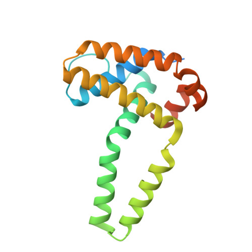
The prosurvival and proapoptotic proteins of the BCL-2 family share a similar three-dimensional fold despite their opposing functions. However, many biochemical studies highlight the requirement for conformational changes for the functioning of both types of proteins, although structural data to support such changes remain elusive. Here, we describe the X-ray structure of dimeric BCL-W that reveals a major conformational change involving helices α3 and α4 hinging away from the core of the protein. Biochemical and functional studies reveal that the α4-α5 hinge region is required for dimerization of BCL-W, and functioning of both pro- and antiapoptotic BCL-2 proteins. Hence, this structure reveals a conformational flexibility not seen in previous BCL-2 protein structures and provides insights into how these regulators of apoptosis can change conformation to exert their function.
- Structural Biology Division, The Walter and Eliza Hall Institute of Medical Research, Parkville, Victoria 3052, Australia.
Organizational Affiliation: