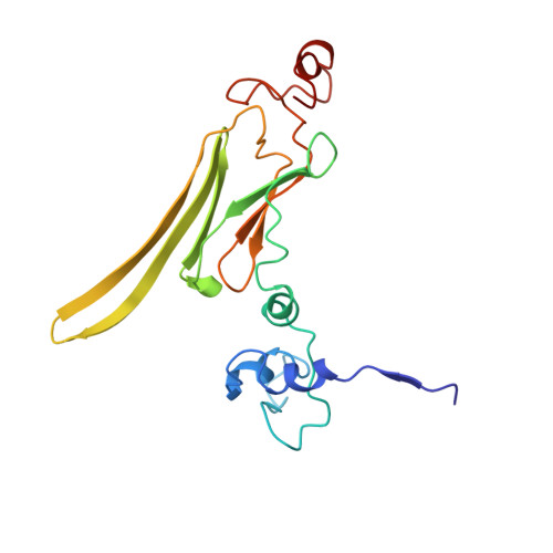
2YGD

Molecular architectures of the 24meric eye lens chaperone alphaB- crystallin elucidated by a triple hybrid approach


Experimental Data Snapshot

  • Method: ELECTRON MICROSCOPY
  • Resolution: 9.40 Å
  • Aggregation State: PARTICLE 
  • Reconstruction Method: SINGLE PARTICLE 

Starting Model: experimental
View more details

wwPDB Validation   3D Report Full Report


This is version 1.4 of the entry. See complete history


Literature

Multiple Molecular Architectures of the Eye Lens Chaperone Alpha Beta-Crystallin Elucidated by a Triple Hybrid Approach

Braun, N.Zacharias, M.Peschek, J.Kastenmueller, A.Zou, J.Hanzlik, M.Haslbeck, M.Rappsilber, J.Buchner, J.Weinkauf, S.

(2011) Proc Natl Acad Sci U S A 108: 20491

  • DOI: https://doi.org/10.1073/pnas.1111014108
  • Primary Citation of Related Structures:  
    2YGD

  • PubMed Abstract: 

    The molecular chaperone αB-crystallin, the major player in maintaining the transparency of the eye lens, prevents stress-damaged and aging lens proteins from aggregation. In nonlenticular cells, it is involved in various neurological diseases, diabetes, and cancer. Given its structural plasticity and dynamics, structure analysis of αB-crystallin presented hitherto a formidable challenge. Here we present a pseudoatomic model of a 24-meric αB-crystallin assembly obtained by a triple hybrid approach combining data from cryoelectron microscopy, NMR spectroscopy, and structural modeling. The model, confirmed by cross-linking and mass spectrometry, shows that the subunits interact within the oligomer in different, defined conformations. We further present the molecular architectures of additional well-defined αB-crystallin assemblies with larger or smaller numbers of subunits, provide the mechanism how "heterogeneity" is achieved by a small set of defined structural variations, and analyze the factors modulating the oligomer equilibrium of αB-crystallin and thus its chaperone activity.


  • Organizational Affiliation

    Center for Integrated Protein Science Munich, Chemistry Department, Technische Universität München, Lichtenbergstrasse 4, 85747 Garching, Germany.


Macromolecules
Find similar proteins by:  (by identity cutoff)  |  3D Structure
Entity ID: 1
MoleculeChains Sequence LengthOrganismDetailsImage
ALPHA-CRYSTALLIN B CHAIN
A, B, C, D, E
A, B, C, D, E, F, G, H, I, J, K, L, M, N, O, P, Q, R, S, T, U, V, W, X
175Homo sapiensMutation(s): 0 
UniProt & NIH Common Fund Data Resources
Find proteins for P02511 (Homo sapiens)
Explore P02511 
Go to UniProtKB:  P02511
PHAROS:  P02511
GTEx:  ENSG00000109846 
Entity Groups  
Sequence Clusters30% Identity50% Identity70% Identity90% Identity95% Identity100% Identity
UniProt GroupP02511
Sequence Annotations
Expand
  • Reference Sequence
Experimental Data & Validation

Experimental Data

  • Method: ELECTRON MICROSCOPY
  • Resolution: 9.40 Å
  • Aggregation State: PARTICLE 
  • Reconstruction Method: SINGLE PARTICLE 
EM Software:
TaskSoftware PackageVersion
RECONSTRUCTIONIMAGIC

Structure Validation

View Full Validation Report


Entry History 

Deposition Data

Revision History  (Full details and data files)

  • Version 1.0: 2011-12-07
    Type: Initial release
  • Version 1.1: 2012-01-11
    Changes: Other
  • Version 1.2: 2013-11-20
    Changes: Source and taxonomy
  • Version 1.3: 2018-10-03
    Changes: Data collection
  • Version 1.4: 2024-05-08
    Changes: Data collection, Database references, Refinement description