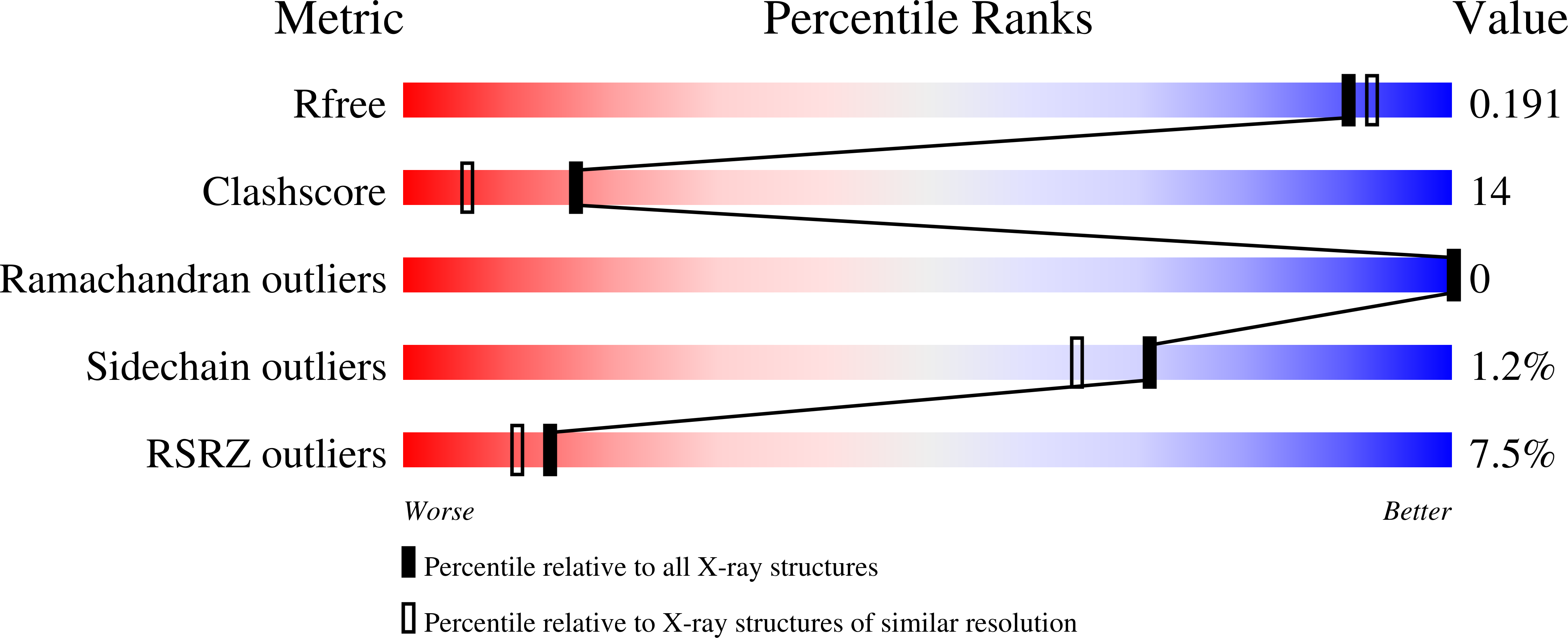
2YHY

Structure of N-Acetylmannosamine kinase in complex with N- acetylmannosamine and ADP


Experimental Data Snapshot

  • Method: X-RAY DIFFRACTION
  • Resolution: 1.82 Å
  • R-Value Free: 0.182 
  • R-Value Work: 0.152 
  • R-Value Observed: 0.154 

Starting Model: experimental
View more details

wwPDB Validation   3D Report Full Report


Ligand Structure Quality Assessment 


This is version 1.3 of the entry. See complete history


Literature

Crystal Structures of N-Acetylmannosamine Kinase Provide Insights Into Enzyme Specificity and Inhibition

Martinez, J.Nguyen, L.D.Tauberger, E.Hinderlich, S.Reutter, W.Fan, H.Saenger, W.Moniot, S.

(2012) J Biol Chem 287: 13656

  • DOI: https://doi.org/10.1074/jbc.M111.318170
  • Primary Citation of Related Structures:  
    2YHW, 2YHY, 2YI1

  • PubMed Abstract: 

    Sialic acids are essential components of membrane glycoconjugates. They are responsible for the interaction, structure, and functionality of all deuterostome cells and have major functions in cellular processes in health and diseases. The key enzyme of the biosynthesis of sialic acid is the bifunctional UDP-N-acetylglucosamine-2-epimerase/N-acetylmannosamine kinase that transforms UDP-N-acetylglucosamine to N-acetylmannosamine (ManNAc) followed by its phosphorylation to ManNAc 6-phosphate and has a direct impact on the sialylation of cell surface components. Here, we present the crystal structures of the human N-acetylmannosamine kinase (MNK) domain of UDP-N-acetylglucosamine-2-epimerase/N-acetylmannosamine kinase in complexes with ManNAc at 1.64 Å resolution, MNK·ManNAc·ADP (1.82 Å) and MNK·ManNAc 6-phosphate · ADP (2.10 Å). Our findings offer detailed insights in the active center of MNK and serve as a structural basis to design inhibitors. We synthesized a novel inhibitor, 6-O-acetyl-ManNAc, which is more potent than those previously tested. Specific inhibitors of sialic acid biosynthesis may serve to further study biological functions of sialic acid.


  • Organizational Affiliation

    From the Institut für Chemie und Biochemie-Kristallographie, Freie Universität Berlin, Takustrasse 6, 14195 Berlin.


Macromolecules
Find similar proteins by:  (by identity cutoff)  |  3D Structure
Entity ID: 1
MoleculeChains Sequence LengthOrganismDetailsImage
BIFUNCTIONAL UDP-N-ACETYLGLUCOSAMINE 2-EPIMERASE/N-ACETYLMANNOSAMINE KINASE343Homo sapiensMutation(s): 0 
EC: 2.7.1.60 (PDB Primary Data), 3.2.1.183 (UniProt)
UniProt & NIH Common Fund Data Resources
Find proteins for Q9Y223 (Homo sapiens)
Explore Q9Y223 
Go to UniProtKB:  Q9Y223
PHAROS:  Q9Y223
GTEx:  ENSG00000159921 
Entity Groups  
Sequence Clusters30% Identity50% Identity70% Identity90% Identity95% Identity100% Identity
UniProt GroupQ9Y223
Sequence Annotations
Expand
  • Reference Sequence
Small Molecules
Ligands 10 Unique
IDChains Name / Formula / InChI Key2D Diagram3D Interactions
ADP
Query on ADP

Download Ideal Coordinates CCD File 
L [auth A]ADENOSINE-5'-DIPHOSPHATE
C10 H15 N5 O10 P2
XTWYTFMLZFPYCI-KQYNXXCUSA-N
2PE
Query on 2PE

Download Ideal Coordinates CCD File 
D [auth A]NONAETHYLENE GLYCOL
C18 H38 O10
YZUUTMGDONTGTN-UHFFFAOYSA-N
BM3
Query on BM3

Download Ideal Coordinates CCD File 
B [auth A]2-acetamido-2-deoxy-alpha-D-mannopyranose
C8 H15 N O6
OVRNDRQMDRJTHS-UOLFYFMNSA-N
PG4
Query on PG4

Download Ideal Coordinates CCD File 
E [auth A]TETRAETHYLENE GLYCOL
C8 H18 O5
UWHCKJMYHZGTIT-UHFFFAOYSA-N
PGE
Query on PGE

Download Ideal Coordinates CCD File 
G [auth A],
J [auth A],
K [auth A]
TRIETHYLENE GLYCOL
C6 H14 O4
ZIBGPFATKBEMQZ-UHFFFAOYSA-N
ZN
Query on ZN

Download Ideal Coordinates CCD File 
O [auth A]ZINC ION
Zn
PTFCDOFLOPIGGS-UHFFFAOYSA-N
EDO
Query on EDO

Download Ideal Coordinates CCD File 
F [auth A],
I [auth A],
M [auth A]
1,2-ETHANEDIOL
C2 H6 O2
LYCAIKOWRPUZTN-UHFFFAOYSA-N
CA
Query on CA

Download Ideal Coordinates CCD File 
C [auth A]CALCIUM ION
Ca
BHPQYMZQTOCNFJ-UHFFFAOYSA-N
CL
Query on CL

Download Ideal Coordinates CCD File 
N [auth A]CHLORIDE ION
Cl
VEXZGXHMUGYJMC-UHFFFAOYSA-M
MG
Query on MG

Download Ideal Coordinates CCD File 
H [auth A]MAGNESIUM ION
Mg
JLVVSXFLKOJNIY-UHFFFAOYSA-N
Binding Affinity Annotations 
IDSourceBinding Affinity
BM3 PDBBind:  2YHY Kd: 2500 (nM) from 1 assay(s)
Experimental Data & Validation

Experimental Data

  • Method: X-RAY DIFFRACTION
  • Resolution: 1.82 Å
  • R-Value Free: 0.182 
  • R-Value Work: 0.152 
  • R-Value Observed: 0.154 
  • Space Group: P 41 21 2
Unit Cell:
Length ( Å )Angle ( ˚ )
a = 90.38α = 90
b = 90.38β = 90
c = 101.16γ = 90
Software Package:
Software NamePurpose
REFMACrefinement
XDSdata reduction
XSCALEdata scaling
PHASERphasing

Structure Validation

View Full Validation Report


Ligand Structure Quality Assessment 


Entry History 

Deposition Data

Revision History  (Full details and data files)

  • Version 1.0: 2012-02-29
    Type: Initial release
  • Version 1.1: 2012-05-02
    Changes: Other
  • Version 1.2: 2020-07-29
    Type: Remediation
    Reason: Carbohydrate remediation
    Changes: Data collection, Derived calculations, Other, Structure summary
  • Version 1.3: 2023-12-20
    Changes: Data collection, Database references, Refinement description, Structure summary