


Crystal structure of D-Alanine:D-Alanine Ligase with ADP and D-Alanine from Thermus thermophius HB8
Kitamura, Y., Yokoyama, S., Kuramitsu, S.To be published.
Experimental Data Snapshot
Starting Model: experimental
View more details
Entity ID: 1 | |||||
|---|---|---|---|---|---|
| Molecule | Chains | Sequence Length | Organism | Details | Image |
| D-alanine--D-alanine ligase | 319 | Thermus thermophilus | Mutation(s): 0 EC: 6.3.2.4 | 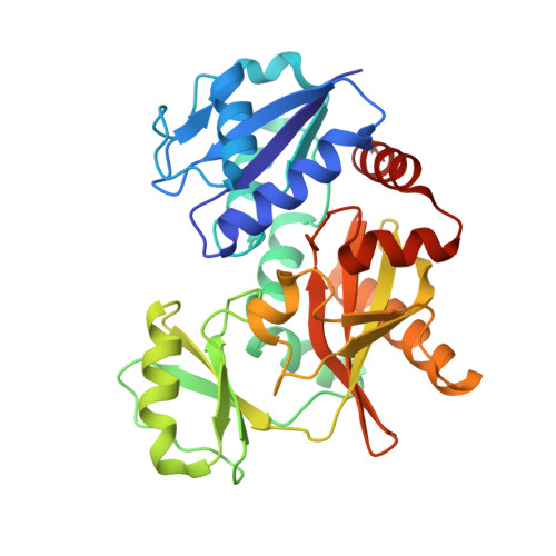 | |
UniProt | |||||
Entity Groups | |||||
| Sequence Clusters | 30% Identity50% Identity70% Identity90% Identity95% Identity100% Identity | ||||
| UniProt Group | Q5SHZ3 | ||||
Sequence AnnotationsExpand | |||||
Reference Sequence | |||||
| Ligands 3 Unique | |||||
|---|---|---|---|---|---|
| ID | Chains | Name / Formula / InChI Key | 2D Diagram | 3D Interactions | |
| ADP Download:Ideal Coordinates CCD File | G [auth A], K [auth B], N [auth C], P [auth D] | ADENOSINE-5'-DIPHOSPHATE C10 H15 N5 O10 P2 XTWYTFMLZFPYCI-KQYNXXCUSA-N |  | ||
| DAL Download:Ideal Coordinates CCD File | H [auth A], Q [auth D] | D-ALANINE C3 H7 N O2 QNAYBMKLOCPYGJ-UWTATZPHSA-N |  | ||
| MG Download:Ideal Coordinates CCD File | E [auth A] F [auth A] I [auth B] J [auth B] L [auth C] | MAGNESIUM ION Mg JLVVSXFLKOJNIY-UHFFFAOYSA-N |  | ||
| Length ( Å ) | Angle ( ˚ ) |
|---|---|
| a = 68.946 | α = 90 |
| b = 105.394 | β = 90 |
| c = 199.748 | γ = 90 |
| Software Name | Purpose |
|---|---|
| CNS | refinement |
| BSS | data collection |
| HKL-2000 | data reduction |
| HKL-2000 | data scaling |
| MOLREP | phasing |


