2ZNV | pdb_00002znv

Crystal structure of human AMSH-LP DUB domain in complex with Lys63-linked ubiquitin dimer


Experimental Data Snapshot

  • Method: X-RAY DIFFRACTION
  • Resolution: 1.60 Å
  • R-Value Free: 
    0.215 (Depositor), 0.215 (DCC) 
  • R-Value Work: 
    0.183 (Depositor), 0.183 (DCC) 
  • R-Value Observed: 
    0.185 (Depositor) 

Starting Models: experimental
View more details

wwPDB Validation 3D Report Full Report

Validation slider image for 2ZNV

This is version 1.4 of the entry. See complete history

Literature

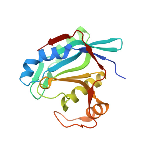
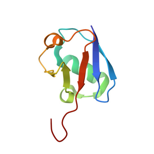
Structural basis for specific cleavage of Lys 63-linked polyubiquitin chains

Sato, Y.Yoshikawa, A.Yamagata, A.Mimura, H.Yamashita, M.Ookata, K.Nureki, O.Iwai, K.Komada, M.Fukai, S.

(2008) Nature 455: 358-362

  • DOI: https://doi.org/10.1038/nature07254
  • Primary Citation Related Structures: 
    2ZNR, 2ZNV

  • PubMed Abstract: 

    Deubiquitinating enzymes (DUBs) remove ubiquitin from conjugated substrates to regulate various cellular processes. The Zn(2+)-dependent DUBs AMSH and AMSH-LP regulate receptor trafficking by specifically cleaving Lys 63-linked polyubiquitin chains from internalized receptors. Here we report the crystal structures of the human AMSH-LP DUB domain alone and in complex with a Lys 63-linked di-ubiquitin at 1.2 A and 1.6 A resolutions, respectively. The AMSH-LP DUB domain consists of a Zn(2+)-coordinating catalytic core and two characteristic insertions, Ins-1 and Ins-2. The distal ubiquitin interacts with Ins-1 and the core, whereas the proximal ubiquitin interacts with Ins-2 and the core. The core and Ins-1 form a catalytic groove that accommodates the Lys 63 side chain of the proximal ubiquitin and the isopeptide-linked carboxy-terminal tail of the distal ubiquitin. This is the first reported structure of a DUB in complex with an isopeptide-linked ubiquitin chain, which reveals the mechanism for Lys 63-linkage-specific deubiquitination by AMSH family members.


  • Organizational Affiliation
    • Structural Biology Laboratory, Life Science Division, Synchrotron Radiation Research Organization and Institute of Molecular and Cellular Biosciences, The University of Tokyo, Tokyo 113-0032, Japan.

Macromolecule Content 

  • Total Structure Weight: 74.74 kDa 
  • Atom Count: 5,691 
  • Modeled Residue Count: 640 
  • Deposited Residue Count: 662 
  • Unique protein chains: 3

Macromolecules

Find similar proteins by:|  3D Structure
Entity ID: 1
MoleculeChains  Sequence LengthOrganismDetailsImage
AMSH-like protease
A, D
178Homo sapiensMutation(s): 1 
EC: 3.1.2.15 (PDB Primary Data), 3.4.19 (UniProt)
UniProt & NIH Common Fund Data Resources
Find proteins for Q96FJ0 (Homo sapiens)
Explore Q96FJ0 
Go to UniProtKB:  Q96FJ0
PHAROS:  Q96FJ0
GTEx:  ENSG00000138134 
Entity Groups
Sequence Clusters30% Identity50% Identity70% Identity90% Identity95% Identity100% Identity
UniProt GroupQ96FJ0
Sequence Annotations
Expand
Reference Sequence
Find similar proteins by:|  3D Structure
Entity ID: 2
MoleculeChains  Sequence LengthOrganismDetailsImage
Ubiquitin
B, E
76Mus musculusMutation(s): 1 
UniProt & NIH Common Fund Data Resources
Find proteins for P0CG50 (Mus musculus)
Explore P0CG50 
Go to UniProtKB:  P0CG50
IMPC:  MGI:98889
Entity Groups
Sequence Clusters30% Identity50% Identity70% Identity90% Identity95% Identity100% Identity
UniProt GroupP0CG50
Sequence Annotations
Expand
Reference Sequence
Find similar proteins by:|  3D Structure
Entity ID: 3
MoleculeChains  Sequence LengthOrganismDetailsImage
Ubiquitin
C, F
77Mus musculusMutation(s): 1 
UniProt & NIH Common Fund Data Resources
Find proteins for P0CG50 (Mus musculus)
Explore P0CG50 
Go to UniProtKB:  P0CG50
IMPC:  MGI:98889
Entity Groups
Sequence Clusters30% Identity50% Identity70% Identity90% Identity95% Identity100% Identity
UniProt GroupP0CG50
Sequence Annotations
Expand
Reference Sequence

Experimental Data & Validation

Experimental Data

  • Method: X-RAY DIFFRACTION
  • Resolution: 1.60 Å
  • R-Value Free:  0.215 (Depositor), 0.215 (DCC) 
  • R-Value Work:  0.183 (Depositor), 0.183 (DCC) 
  • R-Value Observed: 0.185 (Depositor) 
Space Group: P 1 21 1
Unit Cell:
Length ( Å )Angle ( ˚ )
a = 38.089α = 90
b = 97.363β = 97.49
c = 87.894γ = 90
Software Package:
Software NamePurpose
REFMACrefinement
ADSCdata collection
HKL-2000data reduction
HKL-2000data scaling
MOLREPphasing

Structure Validation

View Full Validation Report


Entry History 

Deposition Data

Revision History  (Full details and data files)

  • Version 1.0: 2008-09-02
    Type: Initial release
  • Version 1.1: 2011-07-13
    Changes: Version format compliance
  • Version 1.2: 2021-11-10
    Changes: Database references, Derived calculations
  • Version 1.3: 2023-11-01
    Changes: Data collection, Refinement description
  • Version 1.4: 2024-10-30
    Changes: Structure summary