2ZVT | pdb_00002zvt

Cys285Ser mutant PPARgamma ligand-binding domain complexed with 15-deoxy-delta12,14-prostaglandin J2


Experimental Data Snapshot

  • Method: X-RAY DIFFRACTION
  • Resolution: 1.90 Å
  • R-Value Free: 
    0.275 (Depositor), 0.273 (DCC) 
  • R-Value Work: 
    0.251 (Depositor), 0.249 (DCC) 
  • R-Value Observed: 
    0.251 (Depositor) 

Starting Model: experimental
View more details

wwPDB Validation 3D Report Full Report

Validation slider image for 2ZVT

Ligand Structure Quality Assessment 


This is version 1.3 of the entry. See complete history

Literature

Atomic structure of mutant PPARgamma LBD complexed with 15d-PGJ2: novel modulation mechanism of PPARgamma/RXRalpha function by covalently bound ligands

Waku, T.Shiraki, T.Oyama, T.Morikawa, K.

(2009) FEBS Lett 583: 320-324

  • DOI: https://doi.org/10.1016/j.febslet.2008.12.017
  • Primary Citation Related Structures: 
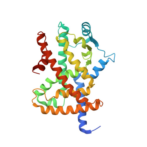
    2ZVT

  • PubMed Abstract: 

    15-deoxy-Delta(12,14)-prostaglandin J(2) (15d-PGJ(2)) activates a nuclear receptor heterodimer, peroxisome proliferators-activated receptor gamma (PPARgamma)/ retinoid X receptor (RXRalpha) through covalent binding to Cys285 in PPARgamma ligand-binding domain (LBD). Here, we present the 1.9A crystal structure of C285S mutant LBD complexed with 15d-PGJ(2), corresponding to the non-covalently bound state. The ligand lies adjacent to a hydrogen-bond network around the helix H2 and the nearby beta-sheet. Comparisons with previous structures clarified the relationships between PPARgamma function and conformational alterations of LBD during the process of covalently binding ligands, such as 15d-PGJ(2), and thus suggested a mechanism, by which these ligands modulate PPARgamma/RXRalpha function through conformational changes of the loop following helix H2' and the beta-sheet.


  • Organizational Affiliation
    • The Takara-Bio Endowed Division, Department of Biomolecular Recognition, Institute for Protein Research, Osaka University, Open Laboratories of Advanced Bioscience and Biotechnology, 6-2-3, Furuedai, Suita, Osaka 565-0874, Japan.

Macromolecule Content 

  • Total Structure Weight: 65.66 kDa 
  • Atom Count: 4,538 
  • Modeled Residue Count: 537 
  • Deposited Residue Count: 572 
  • Unique protein chains: 1

Macromolecules

Find similar proteins by:|  3D Structure
Entity ID: 1
MoleculeChains  Sequence LengthOrganismDetailsImage
Peroxisome proliferator-activated receptor gamma
A, B
286Homo sapiensMutation(s): 1 
Gene Names: PPARG
UniProt & NIH Common Fund Data Resources
Find proteins for P37231 (Homo sapiens)
Explore P37231 
Go to UniProtKB:  P37231
PHAROS:  P37231
GTEx:  ENSG00000132170 
Entity Groups
Sequence Clusters30% Identity50% Identity70% Identity90% Identity95% Identity100% Identity
UniProt GroupP37231
Sequence Annotations
Expand
Reference Sequence

Small Molecules

Experimental Data & Validation

Experimental Data

  • Method: X-RAY DIFFRACTION
  • Resolution: 1.90 Å
  • R-Value Free:  0.275 (Depositor), 0.273 (DCC) 
  • R-Value Work:  0.251 (Depositor), 0.249 (DCC) 
  • R-Value Observed: 0.251 (Depositor) 
Space Group: C 1 2 1
Unit Cell:
Length ( Å )Angle ( ˚ )
a = 92.982α = 90
b = 61.392β = 102.89
c = 118.539γ = 90
Software Package:
Software NamePurpose
CNSrefinement
HKL-2000data collection
HKL-2000data reduction
HKL-2000data scaling
CNSphasing

Structure Validation

View Full Validation Report


Ligand Structure Quality Assessment 


Entry History 

Deposition Data

Revision History  (Full details and data files)

  • Version 1.0: 2009-10-06
    Type: Initial release
  • Version 1.1: 2011-07-13
    Changes: Version format compliance
  • Version 1.2: 2021-11-10
    Changes: Database references, Derived calculations
  • Version 1.3: 2023-11-01
    Changes: Data collection, Refinement description