2ZXZ

Crystal structure of the human RXR alpha ligand binding domain bound to a synthetic agonist compound and a coactivator peptide


Experimental Data Snapshot

  • Method: X-RAY DIFFRACTION
  • Resolution: 3.00 Å
  • R-Value Free: 
    0.278 (Depositor) 
  • R-Value Work: 
    0.206 (Depositor), 0.210 (DCC) 
  • R-Value Observed: 
    0.210 (Depositor) 

Starting Model: experimental
View more details

wwPDB Validation   3D Report Full Report


Ligand Structure Quality Assessment 

Created with Raphaël 2.3.0Worse 01 BetterLigand structure goodness of fit to experimental dataBest fitted P26Click on this verticalbar to view details

This is version 1.3 of the entry. See complete history


Literature

Silicon analogues of the RXR-selective retinoid agonist SR11237 (BMS649): chemistry and biology

Lippert, W.P.Burschka, C.Gotz, K.Kaupp, M.Ivanova, D.Gaudon, C.Sato, Y.Antony, P.Rochel, N.Moras, D.Gronemeyer, H.Tacke, R.

(2009) ChemMedChem 4: 1143-1152

  • DOI: https://doi.org/10.1002/cmdc.200900090
  • Primary Citation of Related Structures:  
    2ZXZ, 2ZY0

  • PubMed Abstract: 

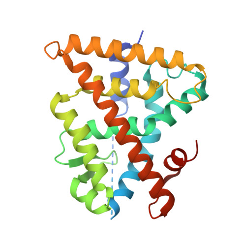
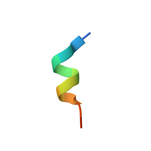
    C/Si switch: Twofold sila-substitution (C/Si exchange) in the RXR-selective retinoids 4 a (SR11237) and 5 a leads to 4 b (disila-SR11237) and 5 b, respectively. Chemistry and biology of the C/Si pairs are reported.SR11237 (BMS649, 4 a) is a pan-RXR-selective retinoid agonist. Its silicon analogue, disila-SR11237 (4 b; twofold C/Si exchange), was prepared in a multistep synthesis by starting from 1,2-bis(ethynyldimethylsilyl)ethane. In addition, the related C/Si analogues 5 a and 5 b, with an indane (disila-indane) instead of a tetraline (disila-tetraline) skeleton, were synthesized. The C/Si pairs 4 a/4 b and 5 a/5 b were studied for their interaction with retinoid receptors and were demonstrated to be highly potent RXR-selective ("rexinoid") agonists. Interestingly, twofold C/Si exchange in the indane moiety of 5 a resulted in a 10-fold increase in biological activity of the corresponding silicon-containing rexinoid 5 b, possibly resulting from an increased receptor affinity or a divergent allosteric effect on co-regulator-binding surfaces. The crystal structures of the ternary complexes formed by 5 a and 5 b, respectively, with the ligand-binding domain of hRXRalpha and a peptide of the co-activator TIF2/GRIP1 revealed additional interactions of the disila analogue 5 b with the H7 and H11 residues, supporting the first option of increased binding affinity. This is the first demonstration of an increase in binding affinity of a ligand to a nuclear receptor by C/Si replacement, thereby adding this C/Si switch strategy to the repertoire of nuclear receptor ligand design.


  • Organizational Affiliation

    Universität Würzburg, Institut für Anorganische Chemie, Am Hubland, 97074 Würzburg, Germany.


Macromolecules
Find similar proteins by:  (by identity cutoff)  |  3D Structure
Entity ID: 1
MoleculeChains Sequence LengthOrganismDetailsImage
Retinoic acid receptor RXR-alpha240Homo sapiensMutation(s): 0 
UniProt & NIH Common Fund Data Resources
Find proteins for P19793 (Homo sapiens)
Explore P19793 
Go to UniProtKB:  P19793
PHAROS:  P19793
GTEx:  ENSG00000186350 
Entity Groups  
Sequence Clusters30% Identity50% Identity70% Identity90% Identity95% Identity100% Identity
UniProt GroupP19793
Sequence Annotations
Expand
  • Reference Sequence

Find similar proteins by:  Sequence   |   3D Structure  

Entity ID: 2
MoleculeChains Sequence LengthOrganismDetailsImage
GRIP1 from Nuclear receptor coactivator 213N/AMutation(s): 0 
UniProt & NIH Common Fund Data Resources
Find proteins for Q15596 (Homo sapiens)
Explore Q15596 
Go to UniProtKB:  Q15596
PHAROS:  Q15596
GTEx:  ENSG00000140396 
Entity Groups  
Sequence Clusters30% Identity50% Identity70% Identity90% Identity95% Identity100% Identity
UniProt GroupQ15596
Sequence Annotations
Expand
  • Reference Sequence
Small Molecules
Ligands 1 Unique
IDChains Name / Formula / InChI Key2D Diagram3D Interactions
P26
Query on P26

Download Ideal Coordinates CCD File 
C [auth A]4-[2-(1,1,3,3-tetramethyl-2,3-dihydro-1H-inden-5-yl)-1,3-dioxolan-2-yl]benzoic acid
C23 H26 O4
ISZBCQLHSNHWLM-UHFFFAOYSA-N
Experimental Data & Validation

Experimental Data

  • Method: X-RAY DIFFRACTION
  • Resolution: 3.00 Å
  • R-Value Free:  0.278 (Depositor) 
  • R-Value Work:  0.206 (Depositor), 0.210 (DCC) 
  • R-Value Observed: 0.210 (Depositor) 
Space Group: P 43 21 2
Unit Cell:
Length ( Å )Angle ( ˚ )
a = 64.079α = 90
b = 64.079β = 90
c = 110.389γ = 90
Software Package:
Software NamePurpose
DENZOdata reduction
SCALEPACKdata scaling
REFMACrefinement
PDB_EXTRACTdata extraction
HKL-2000data reduction
HKL-2000data scaling
AMoREphasing

Structure Validation

View Full Validation Report


Ligand Structure Quality Assessment 

Created with Raphaël 2.3.0Worse 01 BetterLigand structure goodness of fit to experimental dataBest fitted P26Click on this verticalbar to view details

Entry History 

Deposition Data

Revision History  (Full details and data files)

  • Version 1.0: 2009-08-11
    Type: Initial release
  • Version 1.1: 2011-07-13
    Changes: Version format compliance
  • Version 1.2: 2017-10-11
    Changes: Advisory, Refinement description
  • Version 1.3: 2023-11-01
    Changes: Advisory, Data collection, Database references, Derived calculations, Refinement description