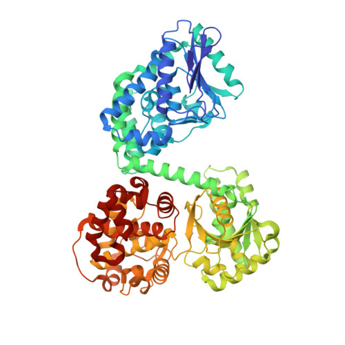
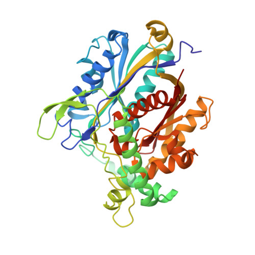
2D3T

Fatty Acid beta-oxidation multienzyme complex from Pseudomonas Fragi, Form V


Experimental Data Snapshot

  • Method: X-RAY DIFFRACTION
  • Resolution: 3.40 Å
  • R-Value Free: 0.295 
  • R-Value Work: 0.242 
  • R-Value Observed: 0.248 

Starting Model: experimental
View more details

wwPDB Validation   3D Report Full Report


Ligand Structure Quality Assessment 


This is version 1.3 of the entry. See complete history


Literature

Ligand-Induced Domain Rearrangement of Fatty Acid beta-Oxidation Multienzyme Complex

Tsuchiya, D.Shimizu, N.Ishikawa, M.Suzuki, Y.Morikawa, K.

(2006) Structure 14: 237-246

  • DOI: https://doi.org/10.1016/j.str.2005.10.011
  • Primary Citation of Related Structures:  
    2D3T

  • PubMed Abstract: 

    The quaternary structure of a fatty acid beta-oxidation multienzyme complex, catalyzing three sequential reactions, was investigated by X-ray crystallographic and small-angle X-ray solution scattering analyses. X-ray crystallography revealed an intermediate structure of the complex among the previously reported structures. However, the theoretical scattering curves calculated from the crystal structures remarkably disagree with the experimental profiles. Instead, an ensemble of the atomic models, which were all calculated by rigid-body optimization, reasonably explained the experimental data. These structures significantly differ from those in the crystals, but they maintain the substrate binding pocket at the domain boundary. Comparisons among these structures indicated that binding of 3-hydroxyhexadecanoyl-CoA or nicotinamide adenine dinucleotide induces domain rearrangements in the complex. The conformational changes suggest the structural events occurring during the chain reaction catalyzed by the multienzyme complex.


  • Organizational Affiliation

    Biomolecular Engineering Research Institute, 6-2-3 Furuedai, Suita, Osaka 565-0874, Japan.


Macromolecules
Find similar proteins by:  (by identity cutoff)  |  3D Structure
Entity ID: 1
MoleculeChains Sequence LengthOrganismDetailsImage
Fatty oxidation complex alpha subunit
A, B
715Pseudomonas fragiMutation(s): 0 
EC: 4.2.1.17 (PDB Primary Data), 5.3.3.8 (PDB Primary Data), 1.1.1.35 (PDB Primary Data), 5.1.2.3 (PDB Primary Data)
UniProt
Find proteins for P28793 (Pseudomonas fragi)
Explore P28793 
Go to UniProtKB:  P28793
Entity Groups  
Sequence Clusters30% Identity50% Identity70% Identity90% Identity95% Identity100% Identity
UniProt GroupP28793
Sequence Annotations
Expand
  • Reference Sequence
Find similar proteins by:  (by identity cutoff)  |  3D Structure
Entity ID: 2
MoleculeChains Sequence LengthOrganismDetailsImage
3-ketoacyl-CoA thiolase
C, D
390Pseudomonas fragiMutation(s): 0 
EC: 2.3.1.16
UniProt
Find proteins for P28790 (Pseudomonas fragi)
Explore P28790 
Go to UniProtKB:  P28790
Entity Groups  
Sequence Clusters30% Identity50% Identity70% Identity90% Identity95% Identity100% Identity
UniProt GroupP28790
Sequence Annotations
Expand
  • Reference Sequence
Experimental Data & Validation

Experimental Data

  • Method: X-RAY DIFFRACTION
  • Resolution: 3.40 Å
  • R-Value Free: 0.295 
  • R-Value Work: 0.242 
  • R-Value Observed: 0.248 
  • Space Group: P 21 21 21
Unit Cell:
Length ( Å )Angle ( ˚ )
a = 95.227α = 90
b = 137.552β = 90
c = 198.139γ = 90
Software Package:
Software NamePurpose
DENZOdata reduction
SCALEPACKdata scaling
AMoREphasing
CNSrefinement

Structure Validation

View Full Validation Report


Ligand Structure Quality Assessment 


Entry History 

Deposition Data

Revision History  (Full details and data files)

  • Version 1.0: 2006-02-21
    Type: Initial release
  • Version 1.1: 2008-04-30
    Changes: Version format compliance
  • Version 1.2: 2011-07-13
    Changes: Version format compliance
  • Version 1.3: 2023-10-25
    Changes: Data collection, Database references, Derived calculations, Refinement description