2X5S

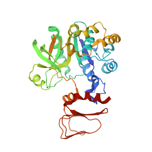
Crystal structure of T. maritima GDP-mannose pyrophosphorylase in apo state.


Experimental Data Snapshot

  • Method: X-RAY DIFFRACTION
  • Resolution: 2.35 Å
  • R-Value Free: 0.273 
  • R-Value Work: 0.224 
  • R-Value Observed: 0.226 

Starting Model: experimental
View more details

wwPDB Validation   3D Report Full Report


This is version 1.4 of the entry. See complete history


Literature

Structural Insights Into the Catalytic Mechanism of Bacterial Guanosine-Diphospho-D-Mannose Pyrophosphorylase and its Regulation by Divalent Ions.

Pelissier, M.C.Lesley, S.Kuhn, P.Bourne, Y.

(2010) J Biol Chem 285: 27468

  • DOI: https://doi.org/10.1074/jbc.M109.095182
  • Primary Citation of Related Structures:  
    2X5S, 2X5Z, 2X60, 2X65

  • PubMed Abstract: 

    GMP catalyzes the formation of GDP-Man, a fundamental precursor for protein glycosylation and bacterial cell wall and capsular polysaccharide biosynthesis. Crystal structures of GMP from the thermophilic bacterium Thermotoga maritima in the apo form, in complex with the substrates mannose-1-phosphate or GTP and bound with the end product GDP-Man in the presence of the essential divalent cation Mg(2+), were solved in the 2.1-2.8 A resolution range. The T. maritima GMP molecule is organized in two separate domains: a N-terminal Rossman fold-like domain and a C-terminal left-handed beta-helix domain. Two molecules associate into a dimer through a tail-to-tail arrangement of the C-terminal domains. Comparative analysis of the structures along with characterization of enzymatic parameters reveals the bases of substrate specificity of this class of sugar nucleotidyltransferases. In particular, substrate and product binding are associated with significant changes in the conformation of loop regions lining the active center and in the relative orientation of the two domains. Involvement of both the N- and C-terminal domains, coupled to the catalytic role of a bivalent metal ion, highlights the catalytic features of bacterial GMPs compared with other members of the pyrophosphorylase superfamily.


  • Organizational Affiliation

    Architecture et Fonction des Macromolécules Biologiques, UMR-6098, CNRS, Université Aix-Marseille, F-13288 Marseille, France.


Macromolecules
Find similar proteins by:  (by identity cutoff)  |  3D Structure
Entity ID: 1
MoleculeChains Sequence LengthOrganismDetailsImage
MANNOSE-1-PHOSPHATE GUANYLYLTRANSFERASE
A, B
336Thermotoga maritima MSB8Mutation(s): 2 
EC: 2.7.7.13
UniProt
Find proteins for Q9X0C3 (Thermotoga maritima (strain ATCC 43589 / DSM 3109 / JCM 10099 / NBRC 100826 / MSB8))
Explore Q9X0C3 
Go to UniProtKB:  Q9X0C3
Entity Groups  
Sequence Clusters30% Identity50% Identity70% Identity90% Identity95% Identity100% Identity
UniProt GroupQ9X0C3
Sequence Annotations
Expand
  • Reference Sequence
Experimental Data & Validation

Experimental Data

  • Method: X-RAY DIFFRACTION
  • Resolution: 2.35 Å
  • R-Value Free: 0.273 
  • R-Value Work: 0.224 
  • R-Value Observed: 0.226 
  • Space Group: P 1 21 1
Unit Cell:
Length ( Å )Angle ( ˚ )
a = 64.011α = 90
b = 92.999β = 110.25
c = 69.687γ = 90
Software Package:
Software NamePurpose
REFMACrefinement
MOSFLMdata reduction
SCALAdata scaling
PHASERphasing

Structure Validation

View Full Validation Report


Entry History 

Deposition Data

Revision History  (Full details and data files)

  • Version 1.0: 2010-06-23
    Type: Initial release
  • Version 1.1: 2011-05-08
    Changes: Version format compliance
  • Version 1.2: 2011-07-13
    Changes: Version format compliance
  • Version 1.3: 2019-10-16
    Changes: Data collection, Experimental preparation, Other
  • Version 1.4: 2023-12-20
    Changes: Data collection, Database references, Derived calculations, Refinement description