3A58

Crystal structure of Sec3p - Rho1p complex from Saccharomyces cerevisiae


Experimental Data Snapshot

  • Method: X-RAY DIFFRACTION
  • Resolution: 2.60 Å
  • R-Value Free: 
    0.265 (Depositor), 0.260 (DCC) 
  • R-Value Work: 
    0.234 (Depositor), 0.230 (DCC) 
  • R-Value Observed: 
    0.235 (Depositor) 

wwPDB Validation   3D Report Full Report


Ligand Structure Quality Assessment 

Created with Raphaël 2.3.0Worse 01 BetterLigand structure goodness of fit to experimental dataBest fitted GNPClick on this verticalbar to view details

This is version 1.4 of the entry. See complete history


Literature

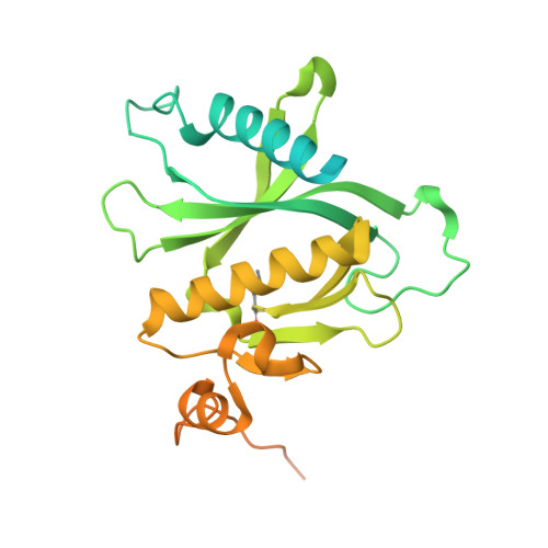
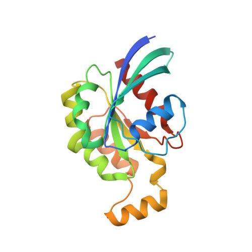
Structural basis for the Rho- and phosphoinositide-dependent localization of the exocyst subunit Sec3

Yamashita, M.Kurokawa, K.Sato, Y.Yamagata, A.Mimura, H.Yoshikawa, A.Sato, K.Nakano, A.Fukai, S.

(2010) Nat Struct Mol Biol 17: 180-186

  • DOI: https://doi.org/10.1038/nsmb.1722
  • Primary Citation of Related Structures:  
    3A58

  • PubMed Abstract: 

    The exocyst complex is a hetero-octameric protein complex that functions during cell polarization by tethering the secretory vesicle to the target membrane. The yeast exocyst subunit Sec3 binds to phosphatidylinositol 4,5-bisphosphate (PtdIns(4,5)P(2)) and the small GTPases Rho1 and Cdc42 via its N-terminal domain (Sec3-N), and these interactions target Sec3 to the plasma membrane. Here we report the crystal structure of the Sec3-N in complex with Rho1 at 2.6-A resolution. Sec3-N adopts a pleckstrin homology (PH) fold, despite having no detectable sequence homology with other PH domains of known structure. Clusters of conserved basic residues constitute a positively charged cleft, which was identified as a binding site for PtdIns(4,5)P(2). Residues Phe77, Ile115 and Leu131 of Sec3 bind to an extended hydrophobic surface formed around switch regions I and II of Rho1. To our knowledge, these are the first structural insights into how an exocyst subunit might interact with both protein and phospholipid factors on the target membrane.


  • Organizational Affiliation

    Structural Biology Laboratory, Life Science Division, Synchrotron Radiation Research Organization and Institute of Molecular and Cellular Biosciences, The University of Tokyo, Tokyo, Japan.


Macromolecules
Find similar proteins by:  (by identity cutoff)  |  3D Structure
Entity ID: 1
MoleculeChains Sequence LengthOrganismDetailsImage
Exocyst complex component SEC3
A, C, E
320Saccharomyces cerevisiaeMutation(s): 0 
Gene Names: SEC3PSL1YER008C
UniProt
Find proteins for P33332 (Saccharomyces cerevisiae (strain ATCC 204508 / S288c))
Explore P33332 
Go to UniProtKB:  P33332
Entity Groups  
Sequence Clusters30% Identity50% Identity70% Identity90% Identity95% Identity100% Identity
UniProt GroupP33332
Sequence Annotations
Expand
  • Reference Sequence
Find similar proteins by:  (by identity cutoff)  |  3D Structure
Entity ID: 2
MoleculeChains Sequence LengthOrganismDetailsImage
GTP-binding protein RHO1
B, D, F
188Saccharomyces cerevisiaeMutation(s): 1 
Gene Names: RHO1YPR165WP9325.3
EC: 3.6.5.2
UniProt
Find proteins for P06780 (Saccharomyces cerevisiae (strain ATCC 204508 / S288c))
Explore P06780 
Go to UniProtKB:  P06780
Entity Groups  
Sequence Clusters30% Identity50% Identity70% Identity90% Identity95% Identity100% Identity
UniProt GroupP06780
Sequence Annotations
Expand
  • Reference Sequence
Small Molecules
Ligands 3 Unique
IDChains Name / Formula / InChI Key2D Diagram3D Interactions
GNP
Query on GNP

Download Ideal Coordinates CCD File 
J [auth B],
O [auth D],
R [auth F]
PHOSPHOAMINOPHOSPHONIC ACID-GUANYLATE ESTER
C10 H17 N6 O13 P3
UQABYHGXWYXDTK-UUOKFMHZSA-N
PO4
Query on PO4

Download Ideal Coordinates CCD File 
G [auth A]
H [auth A]
I [auth A]
L [auth C]
M [auth C]
PHOSPHATE ION
O4 P
NBIIXXVUZAFLBC-UHFFFAOYSA-K
MG
Query on MG

Download Ideal Coordinates CCD File 
K [auth B],
P [auth D],
S [auth F]
MAGNESIUM ION
Mg
JLVVSXFLKOJNIY-UHFFFAOYSA-N
Modified Residues  1 Unique
IDChains TypeFormula2D DiagramParent
MSE
Query on MSE
A, C, E
L-PEPTIDE LINKINGC5 H11 N O2 SeMET
Experimental Data & Validation

Experimental Data

  • Method: X-RAY DIFFRACTION
  • Resolution: 2.60 Å
  • R-Value Free:  0.265 (Depositor), 0.260 (DCC) 
  • R-Value Work:  0.234 (Depositor), 0.230 (DCC) 
  • R-Value Observed: 0.235 (Depositor) 
Space Group: P 41 21 2
Unit Cell:
Length ( Å )Angle ( ˚ )
a = 116α = 90
b = 116.054β = 90
c = 247.675γ = 90
Software Package:
Software NamePurpose
ADSCdata collection
SHARPphasing
CNSrefinement
HKL-2000data reduction
HKL-2000data scaling

Structure Validation

View Full Validation Report


Ligand Structure Quality Assessment 

Created with Raphaël 2.3.0Worse 01 BetterLigand structure goodness of fit to experimental dataBest fitted GNPClick on this verticalbar to view details

Entry History 

Deposition Data

Revision History  (Full details and data files)

  • Version 1.0: 2010-01-12
    Type: Initial release
  • Version 1.1: 2011-07-13
    Changes: Version format compliance
  • Version 1.2: 2013-09-18
    Changes: Derived calculations
  • Version 1.3: 2021-11-10
    Changes: Database references, Derived calculations
  • Version 1.4: 2024-10-23
    Changes: Data collection, Structure summary