3AG9 | pdb_00003ag9

Complex of PKA with the bisubstrate protein kinase inhibitor ARC-1012


Experimental Data Snapshot

  • Method: X-RAY DIFFRACTION
  • Resolution: 2.00 Å
  • R-Value Free: 
    0.246 (Depositor) 
  • R-Value Work: 
    0.201 (Depositor) 
  • R-Value Observed: 
    0.204 (Depositor) 

Starting Model: experimental
View more details

wwPDB Validation 3D Report Full Report

Validation slider image for 3AG9

Ligand Structure Quality Assessment 


This is version 1.4 of the entry. See complete history

Literature

Diversity of bisubstrate binding modes of adenosine analogue-oligoarginine conjugates in protein kinase a and implications for protein substrate interactions.

Pflug, A.Rogozina, J.Lavogina, D.Enkvist, E.Uri, A.Engh, R.A.Bossemeyer, D.

(2010) J Mol Biology 403: 66-77

  • DOI: https://doi.org/10.1016/j.jmb.2010.08.028
  • Primary Citation Related Structures: 
    3AG9, 3AGL, 3AGM

  • PubMed Abstract: 

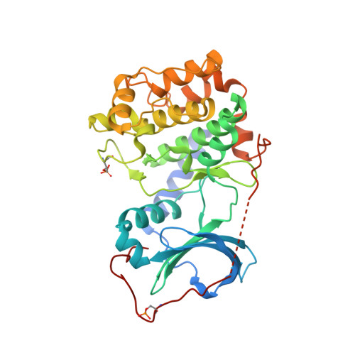
    Crystal structures of the catalytic subunit α of cAMP-dependent protein kinase (PKAc) with three adenosine analogue-oligoarginine conjugates (ARCs) are presented. The rationally designed ARCs include moieties that, in combination, target both the ATP- and the peptide-substrate-binding sites of PKAc, thereby taking advantage of high-affinity binding interactions offered by the ATP site while utilizing an additional mechanism for target specificity via binding to the peptide substrate site. The crystal structures demonstrate that, in accord with the previously reported bisubstrate character of ARCs, the inhibitors occupy both binding sites of PKAc. Further, they show new binding modes that may also apply to natural protein substrates of PKAc, which have not been revealed by previous crystallographic studies. The crystal structures described here contribute to the understanding of the substrate-binding patterns of PKAc and should also facilitate the design of inhibitors targeting PKAc and related protein kinases.


  • Organizational Affiliation
    • Norwegian Structural Biology Centre, Department of Chemistry, University of Tromsø, N-9037 Tromsø, Norway. Electronic address: alexander.pflug@uit.no.

Macromolecule Content 

  • Total Structure Weight: 82.62 kDa 
  • Atom Count: 5,639 
  • Modeled Residue Count: 644 
  • Deposited Residue Count: 702 
  • Unique protein chains: 1

Macromolecules

Find similar proteins by:|  3D Structure
Entity ID: 1
MoleculeChains  Sequence LengthOrganismDetailsImage
cAMP-dependent protein kinase catalytic subunit alpha
A, B
351Bos taurusMutation(s): 0 
Gene Names: PRKACA
EC: 2.7.11.11
UniProt
Find proteins for P00517 (Bos taurus)
Explore P00517 
Go to UniProtKB:  P00517
Entity Groups
Sequence Clusters30% Identity50% Identity70% Identity90% Identity95% Identity100% Identity
UniProt GroupP00517
Sequence Annotations
Expand
Reference Sequence

Small Molecules

Ligands 1 Unique
IDChains Name / Formula / InChI Key2D Diagram3D Interactions
A02

Query on A02


Download:Ideal Coordinates CCD File
C [auth B](10R,20R,23R)-10-(4-aminobutyl)-1-[(2S,3S,4R,5R)-5-(6-amino-9H-purin-9-yl)-3,4-dihydroxytetrahydrofuran-2-yl]-20,23-bis(3-carbamimidamidopropyl)-1,8,11,18,21-pentaoxo-2,9,12,19,22-pentaazatetracosan-24-amide
C40 H70 N18 O9
LIYNJKNSBUCWCZ-SAPGASOESA-N
Modified Residues  2 Unique
IDChains TypeFormula2D DiagramParent
SEP
Query on SEP
A, B
L-PEPTIDE LINKINGC3 H8 N O6 PSER
TPO
Query on TPO
A, B
L-PEPTIDE LINKINGC4 H10 N O6 PTHR

Biologically Interesting Molecules (External Reference) 

1 Unique

Experimental Data & Validation

Experimental Data

  • Method: X-RAY DIFFRACTION
  • Resolution: 2.00 Å
  • R-Value Free:  0.246 (Depositor) 
  • R-Value Work:  0.201 (Depositor) 
  • R-Value Observed: 0.204 (Depositor) 
Space Group: P 21 21 21
Unit Cell:
Length ( Å )Angle ( ˚ )
a = 74.68α = 90
b = 88.04β = 90
c = 115.07γ = 90
Software Package:
Software NamePurpose
REFMACrefinement

Structure Validation

View Full Validation Report


Ligand Structure Quality Assessment 


Entry History 

Deposition Data

Revision History  (Full details and data files)

  • Version 1.0: 2010-09-01
    Type: Initial release
  • Version 1.1: 2011-07-13
    Changes: Atomic model, Database references, Derived calculations, Non-polymer description, Structure summary, Version format compliance
  • Version 1.2: 2012-12-12
    Changes: Other
  • Version 1.3: 2023-08-30
    Changes: Data collection, Database references, Derived calculations, Refinement description
  • Version 1.4: 2024-11-20
    Changes: Structure summary