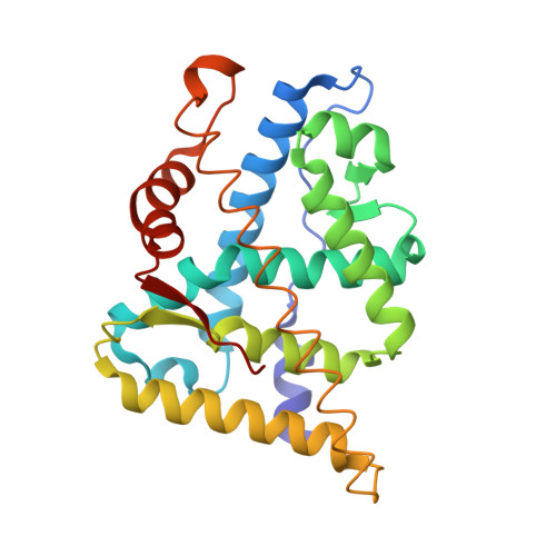
3B65

Crystal structure of the androgen receptor ligand binding domain in complex with SARM S-24


Experimental Data Snapshot

  • Method: X-RAY DIFFRACTION
  • Resolution: 1.80 Å
  • R-Value Free: 0.292 
  • R-Value Work: 0.242 
  • R-Value Observed: 0.242 

wwPDB Validation   3D Report Full Report


Ligand Structure Quality Assessment 

Created with Raphaël 2.3.0Worse 01 BetterLigand structure goodness of fit to experimental dataBest fitted 3B6Click on this verticalbar to view details

This is version 1.2 of the entry. See complete history


Literature

Effect of B-ring substitution pattern on binding mode of propionamide selective androgen receptor modulators

Bohl, C.E.Wu, Z.Chen, J.Mohler, M.L.Yang, J.Hwang, D.J.Mustafa, S.Miller, D.D.Bell, C.E.Dalton, J.T.

(2008) Bioorg Med Chem Lett 18: 5567-5570

  • DOI: https://doi.org/10.1016/j.bmcl.2008.09.002
  • Primary Citation of Related Structures:  
    3B5R, 3B65, 3B66, 3B67, 3B68

  • PubMed Abstract: 

    Selective androgen receptor modulators (SARMs) are essentially prostate sparing androgens, which provide therapeutic potential in osteoporosis, male hormone replacement, and muscle wasting. Herein we report crystal structures of the androgen receptor (AR) ligand-binding domain (LBD) complexed to a series of potent synthetic nonsteroidal SARMs with a substituted pendant arene referred to as the B-ring. We found that hydrophilic B-ring para-substituted analogs exhibit an additional region of hydrogen bonding not seen with steroidal compounds and that multiple halogen substitutions affect the B-ring conformation and aromatic interactions with Trp741. This information elucidates interactions important for high AR binding affinity and provides new insight for structure-based drug design.


  • Organizational Affiliation

    Division of Pharmaceutics, College of Pharmacy, The Ohio State University, 500 West 12th Avenue, 242 L.M. Parks Hall, Columbus, OH 43210, USA.


Macromolecules
Find similar proteins by:  (by identity cutoff)  |  3D Structure
Entity ID: 1
MoleculeChains Sequence LengthOrganismDetailsImage
Androgen receptor249Homo sapiensMutation(s): 0 
Gene Names: ARDHTRNR3C4
UniProt & NIH Common Fund Data Resources
Find proteins for P10275 (Homo sapiens)
Explore P10275 
Go to UniProtKB:  P10275
PHAROS:  P10275
GTEx:  ENSG00000169083 
Entity Groups  
Sequence Clusters30% Identity50% Identity70% Identity90% Identity95% Identity100% Identity
UniProt GroupP10275
Sequence Annotations
Expand
  • Reference Sequence
Small Molecules
Ligands 1 Unique
IDChains Name / Formula / InChI Key2D Diagram3D Interactions
3B6
Query on 3B6

Download Ideal Coordinates CCD File 
B [auth A](2S)-N-(4-cyano-3-iodophenyl)-3-(4-cyanophenoxy)-2-hydroxy-2-methylpropanamide
C18 H14 I N3 O3
RXSZCFAPSDTELY-SFHVURJKSA-N
Binding Affinity Annotations 
IDSourceBinding Affinity
3B6 BindingDB:  3B65 Ki: 0.54 (nM) from 1 assay(s)
PDBBind:  3B65 Ki: 0.54 (nM) from 1 assay(s)
Experimental Data & Validation

Experimental Data

  • Method: X-RAY DIFFRACTION
  • Resolution: 1.80 Å
  • R-Value Free: 0.292 
  • R-Value Work: 0.242 
  • R-Value Observed: 0.242 
  • Space Group: P 21 21 21
Unit Cell:
Length ( Å )Angle ( ˚ )
a = 54.639α = 90
b = 66.712β = 90
c = 69.042γ = 90
Software Package:
Software NamePurpose
CNSrefinement
CrystalCleardata collection
d*TREKdata reduction
d*TREKdata scaling
CNSphasing

Structure Validation

View Full Validation Report


Ligand Structure Quality Assessment 

Created with Raphaël 2.3.0Worse 01 BetterLigand structure goodness of fit to experimental dataBest fitted 3B6Click on this verticalbar to view details

Entry History 

Deposition Data

Revision History  (Full details and data files)

  • Version 1.0: 2008-09-09
    Type: Initial release
  • Version 1.1: 2011-07-13
    Changes: Version format compliance
  • Version 1.2: 2024-02-21
    Changes: Data collection, Database references, Derived calculations