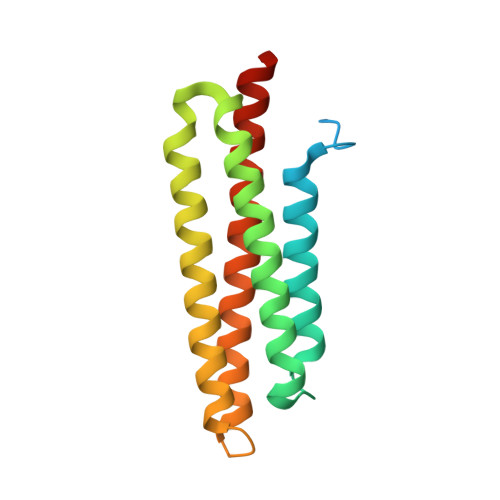
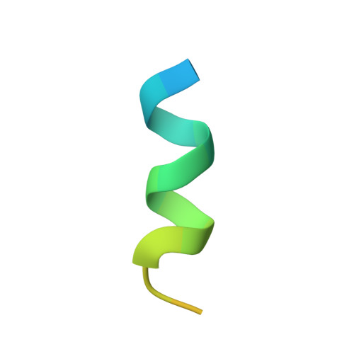
3B71

CD4 endocytosis motif bound to the Focal Adhesion Targeting (FAT) domain of the Focal Adhesion Kinase


Experimental Data Snapshot

  • Method: X-RAY DIFFRACTION
  • Resolution: 2.82 Å
  • R-Value Free: 0.307 
  • R-Value Work: 0.241 
  • R-Value Observed: 0.242 

Starting Model: experimental
View more details

wwPDB Validation   3D Report Full Report


This is version 1.2 of the entry. See complete history


Literature

Structural basis for the interaction between focal adhesion kinase and CD4.

Garron, M.L.Arthos, J.Guichou, J.F.McNally, J.Cicala, C.Arold, S.T.

(2008) J Mol Biol 375: 1320-1328

  • DOI: https://doi.org/10.1016/j.jmb.2007.11.040
  • Primary Citation of Related Structures:  
    3B71

  • PubMed Abstract: 

    Focal adhesion kinase (FAK) and CD4 fulfil vital functions in cellular signal transduction: FAK is a central component in integrin signalling, whereas CD4 plays essential roles in the immune defence. In T lymphocytes, FAK and CD4 localise to the same signalling complexes after stimulation by either the human immunodeficiency virus (HIV) gp120 glycoprotein or an antigen, suggesting the concerted action of FAK and CD4 in these cells. Using crystallography and microcalorimetry, we here show that the focal adhesion targeting (FAT) domain of FAK binds specifically to the CD4 endocytosis motif in vitro. This FAT-CD4 complex is structurally and thermodynamically similar to the one FAT forms with paxillin LD motifs. The CD4 binding site on FAT presents the same features as the established CD4 binding site on the HIV-1 Nef protein. The binding of FAT to CD4 is incompatible with the binding of Lck to CD4. We further show that HIV-1 gp120 triggers the association of CD4 with FAK in T cells, under conditions that are known to dissociate Lck from CD4. Our results suggest that the FAK-CD4 complex represents an alternative route for eliciting T-cell-specific signals and that it links gp120 engagement to distinctive T-cell signalling during HIV infection. In infected cells, HIV-1 Nef may displace FAK from CD4 to protect the cells from apoptosis.


  • Organizational Affiliation

    INSERM, UMR554, 34090 Montpellier, France.


Macromolecules
Find similar proteins by:  (by identity cutoff)  |  3D Structure
Entity ID: 1
MoleculeChains Sequence LengthOrganismDetailsImage
Focal adhesion kinase 1
A, B, C
162Homo sapiensMutation(s): 0 
Gene Names: PTK2FAKFAK1
EC: 2.7.10.2
UniProt & NIH Common Fund Data Resources
Find proteins for Q05397 (Homo sapiens)
Explore Q05397 
Go to UniProtKB:  Q05397
PHAROS:  Q05397
GTEx:  ENSG00000169398 
Entity Groups  
Sequence Clusters30% Identity50% Identity70% Identity90% Identity95% Identity100% Identity
UniProt GroupQ05397
Sequence Annotations
Expand
  • Reference Sequence

Find similar proteins by:  Sequence   |   3D Structure  

Entity ID: 2
MoleculeChains Sequence LengthOrganismDetailsImage
T-cell surface glycoprotein CD4
D, E, F
23Homo sapiensMutation(s): 0 
Gene Names: CD4
UniProt & NIH Common Fund Data Resources
Find proteins for P01730 (Homo sapiens)
Explore P01730 
Go to UniProtKB:  P01730
PHAROS:  P01730
GTEx:  ENSG00000010610 
Entity Groups  
Sequence Clusters30% Identity50% Identity70% Identity90% Identity95% Identity100% Identity
UniProt GroupP01730
Sequence Annotations
Expand
  • Reference Sequence
Experimental Data & Validation

Experimental Data

  • Method: X-RAY DIFFRACTION
  • Resolution: 2.82 Å
  • R-Value Free: 0.307 
  • R-Value Work: 0.241 
  • R-Value Observed: 0.242 
  • Space Group: C 2 2 21
Unit Cell:
Length ( Å )Angle ( ˚ )
a = 88.071α = 90
b = 220.472β = 90
c = 97.546γ = 90
Software Package:
Software NamePurpose
REFMACrefinement
MOSFLMdata reduction
SCALAdata scaling
MOLREPphasing

Structure Validation

View Full Validation Report


Entry History 

Deposition Data

Revision History  (Full details and data files)

  • Version 1.0: 2008-01-01
    Type: Initial release
  • Version 1.1: 2011-07-13
    Changes: Advisory, Version format compliance
  • Version 1.2: 2023-08-30
    Changes: Data collection, Database references, Refinement description