3C5F | pdb_00003c5f

Structure of a binary complex of the R517A Pol lambda mutant


Experimental Data Snapshot

  • Method: X-RAY DIFFRACTION
  • Resolution: 2.25 Å
  • R-Value Free: 
    0.263 (Depositor), 0.252 (DCC) 
  • R-Value Work: 
    0.224 (Depositor), 0.213 (DCC) 

wwPDB Validation 3D Report Full Report

Validation slider image for 3C5F

This is version 1.4 of the entry. See complete history

Literature

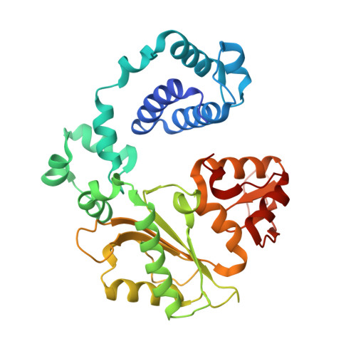
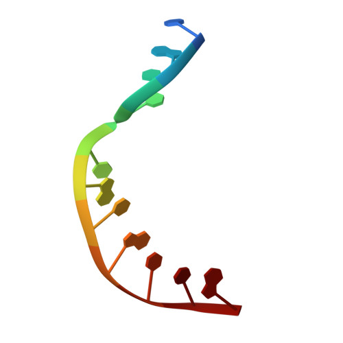
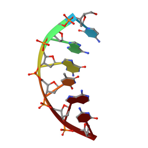
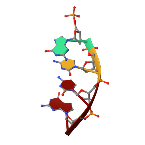
Substrate-induced DNA strand misalignment during catalytic cycling by DNA polymerase lambda.

Bebenek, K.Garcia-Diaz, M.Foley, M.C.Pedersen, L.C.Schlick, T.Kunkel, T.A.

(2008) EMBO Rep 9: 459-464

  • DOI: https://doi.org/10.1038/embor.2008.33
  • Primary Citation Related Structures: 
    3C5F, 3C5G

  • PubMed Abstract: 

    The simple deletion of nucleotides is common in many organisms. It can be advantageous when it activates genes beneficial to microbial survival in adverse environments, and deleterious when it mutates genes relevant to survival, cancer or degenerative diseases. The classical idea is that simple deletions arise by strand slippage. A prime opportunity for slippage occurs during DNA synthesis, but it remains unclear how slippage is controlled during a polymerization cycle. Here, we report crystal structures and molecular dynamics simulations of mutant derivatives of DNA polymerase lambda bound to a primer-template during strand slippage. Relative to the primer strand, the template strand is in multiple conformations, indicating intermediates on the pathway to deletion mutagenesis. Consistent with these intermediates, the mutant polymerases generate single-base deletions at high rates. The results indicate that dNTP-induced template strand repositioning during conformational rearrangements in the catalytic cycle is crucial to controlling the rate of strand slippage.


  • Organizational Affiliation
    • Laboratory of Structural Biology and Laboratory of Molecular Genetics, National Institute of Environmental Health Sciences, NIH, Research Triangle Park, North Carolina 27709, USA.

Macromolecule Content 

  • Total Structure Weight: 87.6 kDa 
  • Atom Count: 6,585 
  • Modeled Residue Count: 694 
  • Deposited Residue Count: 712 
  • Unique protein chains: 1
  • Unique nucleic acid chains: 3

Macromolecules


Find similar proteins by:|  3D Structure
Entity ID: 4
MoleculeChains  Sequence LengthOrganismDetailsImage
DNA polymerase lambdaG [auth A],
H [auth B]
335Homo sapiensMutation(s): 1 
Gene Names: POLL
EC: 2.7.7.7 (PDB Primary Data), 4.2.99 (PDB Primary Data)
UniProt & NIH Common Fund Data Resources
Find proteins for Q9UGP5 (Homo sapiens)
Explore Q9UGP5 
Go to UniProtKB:  Q9UGP5
PHAROS:  Q9UGP5
GTEx:  ENSG00000166169 
Entity Groups
Sequence Clusters30% Identity50% Identity70% Identity90% Identity95% Identity100% Identity
UniProt GroupQ9UGP5
Sequence Annotations
Expand
Reference Sequence
Find similar nucleic acids by:  Sequence
Entity ID: 1
MoleculeChains LengthOrganismImage
DNA (5'-D(*DCP*DGP*DGP*DCP*DCP*DGP*DTP*DAP*DCP*DTP*DG)-3')A [auth T],
D [auth U]
11N/A
Sequence Annotations
Expand
Reference Sequence
Find similar nucleic acids by:  Sequence
Entity ID: 2
MoleculeChains LengthOrganismImage
DNA (5'-D(*DCP*DAP*DGP*DTP*DAP*DC)-3')B [auth P],
E [auth Q]
6N/A
Sequence Annotations
Expand
Reference Sequence
Find similar nucleic acids by:  Sequence
Entity ID: 3
MoleculeChains LengthOrganismImage
DNA (5'-D(P*DGP*DCP*DCP*DG)-3')C [auth D],
F [auth E]
4N/A
Sequence Annotations
Expand
Reference Sequence

Experimental Data & Validation

Experimental Data

  • Method: X-RAY DIFFRACTION
  • Resolution: 2.25 Å
  • R-Value Free:  0.263 (Depositor), 0.252 (DCC) 
  • R-Value Work:  0.224 (Depositor), 0.213 (DCC) 
Space Group: P 21 21 2
Unit Cell:
Length ( Å )Angle ( ˚ )
a = 94.237α = 90
b = 151.884β = 90
c = 85.635γ = 90
Software Package:
Software NamePurpose
DENZOdata reduction
SCALEPACKdata scaling
CNSrefinement
PDB_EXTRACTdata extraction
HKL-2000data collection
CNSphasing

Structure Validation

View Full Validation Report


Entry History 

Deposition Data

Revision History  (Full details and data files)

  • Version 1.0: 2008-09-02
    Type: Initial release
  • Version 1.1: 2011-07-13
    Changes: Version format compliance
  • Version 1.2: 2017-10-25
    Changes: Refinement description
  • Version 1.3: 2021-10-20
    Changes: Database references, Derived calculations
  • Version 1.4: 2024-10-30
    Changes: Data collection, Structure summary