The dimer interface of the membrane type 1 matrix metalloproteinase hemopexin domain: crystal structure and biological functions
Tochowicz, A., Goettig, P., Evans, R., Visse, R., Shitomi, Y., Palmisano, R., Ito, N., Richter, K., Maskos, K., Franke, D., Svergun, D., Nagase, H., Bode, W., Itoh, Y.(2011) J Biol Chem 286: 7587-7600
- PubMed: 21193411 Search on PubMedSearch on PubMed Central
- DOI: https://doi.org/10.1074/jbc.M110.178434
- Primary Citation Related Structures:
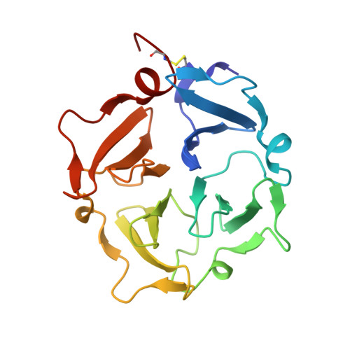
3C7X - PubMed Abstract:
Homodimerization is an essential step for membrane type 1 matrix metalloproteinase (MT1-MMP) to activate proMMP-2 and to degrade collagen on the cell surface. To uncover the molecular basis of the hemopexin (Hpx) domain-driven dimerization of MT1-MMP, a crystal structure of the Hpx domain was solved at 1.7 Å resolution. Two interactions were identified as potential biological dimer interfaces in the crystal structure, and mutagenesis studies revealed that the biological dimer possesses a symmetrical interaction where blades II and III of molecule A interact with blades III and II of molecule B. The mutations of amino acids involved in the interaction weakened the dimer interaction of Hpx domains in solution, and incorporation of these mutations into the full-length enzyme significantly inhibited dimer-dependent functions on the cell surface, including proMMP-2 activation, collagen degradation, and invasion into the three-dimensional collagen matrix, whereas dimer-independent functions, including gelatin film degradation and two-dimensional cell migration, were not affected. These results shed light on the structural basis of MT1-MMP dimerization that is crucial to promote cellular invasion.
- Arbeitsgruppe Proteinaseforschung, Max-Planck-Institut fuer Biochemie, Am Klopferspitz 18, D-82152 Martinsried, Germany.
Organizational Affiliation: